
Human Pancreatic Islets
Cat.No.: CSC-C4882L
Species: Human
Source: Pancreatic Islet; Pancreas
- Specification
- Background
- Scientific Data
- Q & A
- Customer Review
Never can cryopreserved cells be kept at -20 °C.
Human Pancreatic Islets are highly specialized 3-dimensional microorgans isolated from the pancreas of human donors. Instead of a single cell line, they are a heterogeneous population of endocrine cells, mostly insulin-producing β cells, glucagon-secreting α cells, somatostatin-producing δ cells, and pancreatic polypeptide (PP) cells. These cells collectively play a key role in the maintenance of glucose homeostasis through tightly regulated hormone secretion. Human pancreatic islets are one of the most valuable systems for the study of human Diabetes mellitus as they recapitulate closely native pancreatic endocrine function and are physiologically relevant ex vivo models. They are widely used in studies of glucose-stimulated insulin secretion (GSIS), beta-cell dysfunction, and islet transplantation biology. In contrast to immortalized β-cell lines, human islets preserve native cell-cell interactions and intrinsic signaling networks and are more translationally relevant for metabolic and pharmacological studies.
Furthermore, human pancreatic islets are important for the evaluation of drug-induced β-cell toxicity, screening of anti-diabetic compounds, and study of mechanisms of islet inflammation and immune-mediated damage. They are also a vital model in regenerative medicine and cell replacement therapy research. Despite limitations in human pancreatic islet availability and donor variability, they are the gold standard in vitro system for the study of human pancreatic endocrine physiology and diabetes pathophysiology.
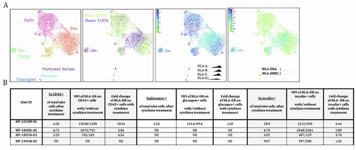
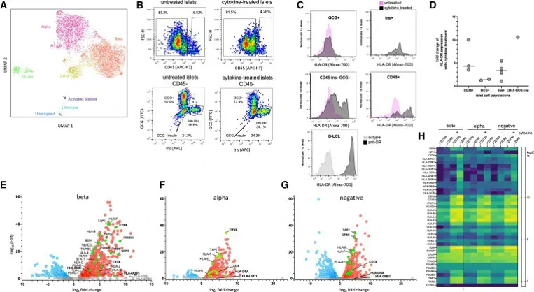
Cytokine Treatment of Cultured Human Islets Upregulates MHC-I and MHC-II Antigen Presentation Pathways
Type 1 diabetes (T1D) involves T cell destruction of β-cells, potentially triggered by inflammatory epitopes. To characterize the human islet immunopeptidome, Nanaware et al. treated cadaveric non-diabetic islets with pro-inflammatory cytokines.
Cytokine treatment induced the loss of acinar cells and significantly upregulated MHC-II (HLA-DR) on insulin-positive and immune cells (Fig.1A-D; Fig. 2B). Bulk RNA-seq of sorted populations revealed the most pronounced transcriptional upregulation of MHC-I (NLRC5, HLA-A/B/C) and MHC-II (CIITA, HLA-DP/DQ) antigen presentation pathways in β-cells (Fig. 1E-H), corroborated by scRNA-seq (Fig. 2A).
Critically, genes upregulated by cytokine treatment overlapped significantly with those identified in β-cells from T1D donors versus controls (p < 0.001), validating the in vitro model against in vivo disease states. These data demonstrate that cytokine stimulation effectively simulates the T1D milieu by enhancing MHC expression and antigen presentation machinery in human islets.
Ask a Question
Write your own review
- You May Also Need

Description: Primary Human Pancreatic Stromal Cells were initiated by elutriation from normal human pancreatic tissue.These cells were originated using Complete Serum-Free Medium Kit With SuperFuel™, are ...

Description: Human Pancreatic Microvascular Endothelial Cells from Creative Bioarray are isolated from human pancreatic tissue. Human Pancreatic Microvascular Endothelial Cells are grown in T25 tissue culture ...
Description: Pancreatic Stellate Cells (HPaSteC) are the main fibroblastic cells of the pancreas. HPaSteC are responsible for the synthesis and the degradation of the extracellular matrix proteins that promote ...
Description: Recent research indicates cancer associated fibroblasts (CAFs) significant involvement in crucial aspects of epithelial solid tumor biology, specifically neoplastic progression, tumor growth, ...

Description: Human Primary Pancreatic Fibroblasts are isolated from normal human pancreatic tissue. Human Primary Pancreatic Fibroblasts are grown in T75 tissue culture flasks pre-coated with gelatin-based ...
Description: Human Pancreatic Epithelial Cells from Creative Bioarray are isolated from normal human pancreatic tissue. Human Pancreatic Epithelial Cells are grown in T25 tissue culture flasks pre-coated with ...