Orthotopic Models
- Background
- Models
- Core Capabilities
- Features
- FAQ
Ultimate Clinical Relevance through Organ-Specific Tumor Modeling
Leveraging our orthotopic platform, we provide high fidelity CDX/PDX models with native TME, advanced longitudinal imaging and IND-ready data packages.
Why Orthotopic Models?
Recapitulating the Complexity of Human Cancer
While subcutaneous models are common, they often fail to predict clinical outcomes due to the lack of organ-specific microenvironments. Our orthotopic models provide:
- Native TME Architecture: Preserves critical stromal, vascular, and immune interactions.
- Clinically Relevant Metastasis: Enables study of spontaneous metastasis to secondary organs (e.g., liver, lung, bone).
- Superior PK/PD Correlation: Mimics human drug distribution and blood-organ barrier challenges (e.g., Blood-Brain Barrier).
Orthotopic vs. Subcutaneous Models
| Feature | Subcutaneous Models | Orthotopic Models (Our Platform) |
|---|---|---|
| Microenvironment | Ectopic (Skin) | Native (Organ-specific) |
| Metastasis | Rarely occurs | High (Spontaneous & Distant) |
| Predictive Value | Moderate | High (Clinically Correlated) |
| Imaging Monitoring | Manual Caliper | Longitudinal (BLI / MRI / Ultrasound) |
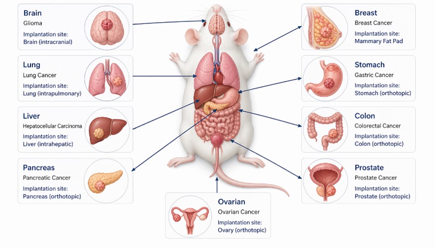
Our Validated Model Portfolio
CNS Tumor Models (Brain & Glioma)
- Indications: Glioblastoma (GBM), medulloblastoma
- Model Types: CDX / PDX
- Key Features: Blood–brain barrier (BBB) relevance, invasive growth patterns
- Applications: Neuro-oncology, CNS drug delivery, targeted therapies
Lung Cancer Models
- Indications: NSCLC, SCLC
- Model Types: CDX / PDX
- Key Features: Orthotopic lung colonization, spontaneous metastasis
- Applications: Anti-tumor efficacy, metastasis studies
Gastrointestinal Tumor Models
- Indications: Colorectal, pancreatic, gastric cancers
- Model Types: CDX / PDX / Syngeneic
- Key Features: Rich stromal environment, high metastatic potential (liver/peritoneum)
- Applications: Drug efficacy, metastasis research, tumor–stroma interaction
Liver Cancer Models
- Indications: Hepatocellular carcinoma (HCC)
- Model Types: CDX / PDX
- Key Features: Native hepatic microenvironment, vascularized tumors
- Applications: Targeted therapy, metabolism-related oncology
Breast Cancer Models
- Indications: TNBC, HER2+, ER+
- Model Types: CDX / PDX
- Key Features: Mammary fat pad implantation, lung metastasis
- Applications: Metastasis studies, drug screening, resistance mechanisms
Urogenital Tumor Models
- Indications: Prostate, bladder cancers
- Model Types: CDX / PDX / Syngeneic
- Key Features: Hormone-responsive environment, local invasion
- Applications: Hormone therapy, immuno-oncology
Core Capabilities

01. Precision Model Engineering
We establish high-fidelity orthotopic tumor models by implanting tumor cells or patient-derived tissues into the corresponding organ site, ensuring physiological relevance and reproducibility.
- Diverse Platform: Expertly validated CDX, PDX, and syngeneic models.
- Organ-specific implantation (brain, lung, liver, pancreas, etc.)
- Superior Reproducibility: High take rates with consistent growth kinetics for robust study design.
- TME Preservation: Maintains critical stromal, vascular, and immune architecture essential for drug response evaluation.
Ensures clinically relevant tumor growth and disease progression.
02. Advanced In Vivo Imaging
We utilize multi-modal imaging technologies to enable non-invasive, real-time monitoring of tumor development and response.
- Bioluminescence imaging (IVIS/BLI)
- Fluorescence imaging
- MRI-based tumor visualization (select models)
- Longitudinal tumor burden quantification
Enables dynamic, high-resolution tracking of tumor progression and metastasis.
03. Drug Efficacy & Translational Research
Our platform supports comprehensive oncology drug evaluation under physiologically relevant conditions, enabling strong translational insights.
- Broad Therapeutic Modalities: Small molecules, biologics, ADCs, and immunotherapies
- Comprehensive Study Design: Monotherapy and combination therapy studies
- Translational Insights: PK/PD and efficacy correlation analysis
- Survival & Metastasis Endpoints: Robust statistical data to support critical go/no-go decision-making.
Provides robust data to support preclinical decision-making and IND-enabling studies.
Why Choose Creative Bioarray?

Strong Expertise in Complex Models
Orthotopic models require advanced surgical and technical capabilities. Our experienced team ensures high success rates, reproducibility, and consistency across studies.

Integrated Imaging & Monitoring
We combine IVIS, fluorescence, and MRI technologies to enable non-invasive, real-time tracking of tumor growth and metastasis.

Flexible & Customized Study Design
Every project is tailored to your needs, including model selection, dosing strategies, and endpoint design, ensuring alignment with your development goals.

Fast Turnaround & Reliable Delivery
Optimized workflows and dedicated project management ensure efficient execution and high-quality data delivery on time.
FAQ
What is the advantage of orthotopic tumor models over subcutaneous models?
Orthotopic models better replicate the tumor microenvironment and metastatic behavior, resulting in higher clinical relevance and improved predictive power.
Which tumor types can be modeled orthotopically?
We support a wide range of tumor types, including brain, lung, breast, liver, pancreatic, colorectal, and urogenital cancers.
Can orthotopic models be used for metastasis studies?
Yes. These models enable spontaneous metastasis, making them ideal for studying tumor progression and anti-metastatic therapies.
What imaging methods are available?
We offer bioluminescence (IVIS), fluorescence imaging, and MRI (select models) for real-time tumor monitoring.
Do you provide customized study design?
Yes. We offer tailored study design, flexible endpoints, and full project support based on your research goals.
Ready to Advance Your Oncology Research?
Leverage our clinically relevant orthotopic tumor models to generate high-quality, decision-ready preclinical data.