Type 1 Diabetes Mellitus Model
Type 1 diabetes is an autoimmune disease characterized by complete insulin deficiency. With the development of economy and the improvement of people's living standard, the morbidity and mortality are increasing year by year. The causative factors of Type 1 diabetes mainly include genetic susceptibility, immunologic dysregulation and exposure to environmental triggers. But the etiology of type 1 diabetes remains unclear. At present, various animal models including spontaneously developing strains, chemical-induced strains and creating diabetes-prone species through gene transfer techniques are used to study Type 1 diabetes.
Creative Bioarray focuses on drug research and development services and helps customers assess/evaluate the drug efficacy and study the associated pathological mechanisms of T1DM by Type 1 diabetes mellitus model.
Type 1 diabetes animal models include but not limited to:
- Chemical-induced Type 1 Diabetes Models
- Transgenic Models
- Surgical Models
Species available
- Rat
- Mouse
- Rabbit
- Dog
- Pig
Our capabilities
- The transparency of the lens was observed using a slit-lamp and anterior segment in both eyes of rat were imaged using an anterior segment camera system.
- We test the levels of advanced glycation end products (AGEs), MDA, GSH as well as the activities of SOD and CAT by Elisa.
Assays available
- Evaluation of progression of cataract
- Biochemical analysis
- Pathological evaluation of lens
With extensive experience in the field of T1DM, we are confident to help you overcome any upcoming challenges. Our experts are fully capable of customizing our protocols and assays to meet your specific needs. With our help, we wish to facilitate your research with high efficiency.
Study examples
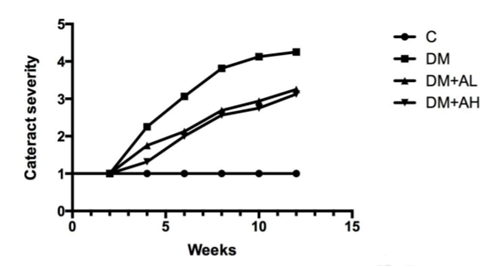 Figure. 1. Quantitive representation of cataract progression in each group with time. Stage of cataract in each group was averaged at a given time.
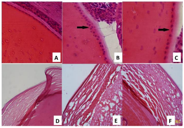 Figure. 2. Hematoxylin-eosin staining of rat lens paraffin sections: (A) organized monolayer of cuboidal lens epithelial cells in the C group (×400); (B) multiple layers of lens epithelial cells can be seen in the DM group (×400); (C) lens epithelial cells are still in a monolayer in the AL group with slight disorganization (×400); (D) lens section from the C group (×200); (E) DM group (×200); (F) DM+AL group (×200).
Quotation and ordering
If you have any special needs or questions regarding our services, please feel free to contact us. We look forward to cooperating with you in the future.
Reference
Yang, Ming, et al. "Effect of astaxanthin on metabolic cataract in rats with type 1 diabetes mellitus." Experimental and Molecular Pathology (2020): 104372.
For research use only. Not for any other purpose.
Disease Models
- Oncology Models
-
Inflammation & Autoimmune Disease Models
- Rheumatoid Arthritis Models
- Glomerulonephritis Models
- Multiple Sclerosis (MS) Models
- Ocular Inflammation Models
- Sjögren's Syndrome Model
- LPS-induced Acute Lung Injury Model
- Peritonitis Models
- Passive Cutaneous Anaphylaxis Model
- Delayed-Type Hypersensitivity (DTH) Models
- Inflammatory Bowel Disease Models
- Systemic Lupus Erythematosus Animal Models
- Oral Mucositis Model
- Asthma Model
- Sepsis Model
- Psoriasis Model
- Atopic Dermatitis (AD) Model
- Scleroderma Model
- Gouty Arthritis Model
- Carrageenan-Induced Air Pouch Synovitis Model
- Carrageenan-Induced Paw Edema Model
- Experimental Autoimmune Myasthenia Gravis (EAMG) Model
- Graft-versus-host Disease (GvHD) Models
-
Cardiovascular Disease Models
- Surgical Models
- Animal Models of Hypertension
- Venous Thrombosis Model
- Atherosclerosis model
- Cardiac Arrhythmia Model
- Hyperlipoidemia Model
- Doxorubicin-induced Heart Failure Model
- Isoproterenol-induced Heart Failure Model
- Arterial Thrombosis Model
- Pulmonary Arterial Hypertension (PAH) Models
- Heart Failure with Preserved Ejection Fraction (HFpEF) Model
- Cardio-Renal-Metabolic (CKM) Syndrome Model
-
Neurological Disease Models
- Alzheimer's Disease Modeling and Assays
- Seizure Models
- Parkinson's Disease Models
- Ischemic Stroke Models
- Acute Spinal Cord Injury (ASCI) Model
- Traumatic Brain Injury (TBI) Model
- Hypoxic-Ischemic Encephalopathy (HIE) Model
- Tourette Syndrome (TS) Model
- Amyotrophic Lateral Sclerosis (ALS) Model
- Huntington's Disease (HD) Model
- Intracerebral hemorrhage (ICH) Models
- Schizophrenia Model
- Depression Models
- Pain Models
-
Metabolic Disease Models
- Type 1 Diabetes Mellitus Model
- Type 2 Diabetes Mellitus Model
- Animal Model of Hyperuricemia
-
Nonalcoholic Fatty Liver Disease Model
- High-Fat Diet-Induced Nonalcoholic Fatty Liver Disease (NAFLD) Model
- Methionine and Choline Deficient (MCD) Diet-Induced Nonalcoholic Fatty Liver Disease (NAFLD) Model
- Gubra-Amylin NASH (GAN) Diet-Induced Nonalcoholic Fatty Liver Disease (NAFLD) Model
- Streptozotocin (STZ) Induced Nonalcoholic Fatty Liver Disease (NAFLD) Model
- High Fat Diet-Induced Obesity Model
- Diabetic Foot Ulcer (DFU) Model
- Cardio-Renal-Metabolic (CKM) Syndrome Model
- Liver Disease Models
- Rare Disease Models
- Respiratory Disease Models
- Digestive Disease Models
-
Urology Disease Models
- Cisplatin-induced Nephrotoxicity Model
- Unilateral Ureteral Obstruction Model
- 5/6 Nephrectomy Model
- Renal Ischemia-Reperfusion Injury (RIRI) Model
- Diabetic Nephropathy (DN) Models
- Passive Heymann Nephritis (PHN) Model
- Adenine-Induced Chronic Kidney Disease (CKD) Model
- Kidney Stone Model
- Doxorubicin-Induced Nephropathy Model
- Orthotopic Kidney Transplantation Model
- Benign Prostatic Hyperplasia (BPH) Model
- Peritoneal Fibrosis Model
- Cardio-Renal-Metabolic (CKM) Syndrome Model
- Orthopedic Disease Models
- Ocular Disease Models
- Infectious Disease Models
- Skin Disease Models
- Otology Disease Models