What Is the Use of FISH in Solid Tumors?
Fluorescence in situ hybridization (FISH) has emerged as a powerful molecular technique in the field of cancer research, particularly in the study of solid tumors. Solid tumors, characterized by the abnormal growth of cells in tissues, represent a significant challenge in terms of diagnosis, treatment, and understanding of their underlying genetic alterations. FISH, with its ability to visualize specific genetic abnormalities within individual cells, has revolutionized our understanding of solid tumors and paved the way for improved diagnosis and targeted therapies.
FISH Techniques Used in Solid Tumor Research
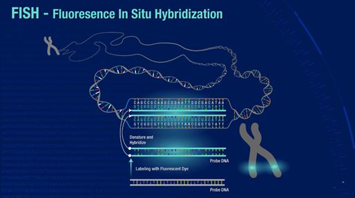
The principles of FISH involve the use of fluorescently labeled DNA probes that hybridize to complementary target sequences within the tumor cells. In solid tumor research, FISH techniques are used to detect various chromosomal aberrations, including gene amplifications, deletions, translocations, and rearrangements. By targeting specific genes or genomic regions of interest, FISH enables the identification of genetic alterations that play crucial roles in tumor development and progression.
FISH techniques used in solid tumor research encompass a variety of methods that allow for the detection and visualization of specific genetic alterations within tumor cells. These techniques play a vital role in understanding the genetic complexities of solid tumors, identifying potential diagnostic and prognostic markers, and guiding treatment decisions.
- DNA probe-based FISH. It is the most widely used technique in solid tumor research. It involves the use of fluorescently labeled DNA probes that hybridize to complementary target sequences within the tumor cells. These probes can be designed to target specific genes, genomic regions, or chromosomal abnormalities associated with solid tumors. By using different fluorescent labels for different probes, it is possible to simultaneously visualize multiple genetic alterations within the tumor cells. This multiplexing capability enhances the efficiency and accuracy of genetic analysis in solid tumors.
- RNA probe-based FISH. It involves the use of fluorescently labeled RNA probes that hybridize to complementary RNA molecules within the tumor cells. These probes are designed to target specific mRNA transcripts or non-coding RNAs of interest. The probes can be labeled with fluorophores that emit distinct colors, enabling the visualization of multiple RNA targets simultaneously.
Prognostic and Predictive Marker Identification
One of the key applications of FISH in solid tumor research is the identification of prognostic and predictive markers. Prognostic markers provide information about the likely outcome of the disease, while predictive markers help in determining the response to specific treatments. By analyzing genetic alterations through FISH, researchers can identify markers associated with disease progression, metastasis, and treatment response.
For instance, in lung cancer, FISH analysis can detect chromosomal rearrangements involving the ALK gene, leading to the identification of patients who are likely to respond to ALK inhibitors. Similarly, in prostate cancer, FISH can detect gene deletions or rearrangements of the androgen receptor (AR) gene, aiding in predicting resistance to hormonal therapies. Such prognostic and predictive markers identified through FISH analysis provide valuable information for personalized treatment decisions.
Evaluation of Treatment Response
FISH techniques play a crucial role in evaluating treatment response in solid tumors. During cancer treatment, genetic alterations within tumor cells can change, leading to the development of resistance or the emergence of new subclones. FISH allows researchers to monitor these changes and assess treatment efficacy.
For example, in breast cancer, FISH analysis can be used to evaluate the response to HER2-targeted therapies by monitoring the status of HER2 gene amplification. Changes in HER2 amplification levels detected by FISH can indicate the development of resistance or response to treatment. By providing real-time information on treatment response, FISH contributes to the optimization of therapeutic strategies and the identification of potential drug resistance mechanisms.
Examples of FISH Applications in Various Solid Tumor Types
FISH has found extensive applications in the study of various solid tumor types, enabling a deeper understanding of their genetic complexities and providing valuable clinical insights. Here are a few examples of FISH applications in specific solid tumors:
- Breast cancer. In breast cancer, FISH analysis is widely used to detect amplifications of genes such as HER2 (human epidermal growth factor receptor 2), indicating the eligibility for HER2-targeted therapies. FISH also plays a role in identifying other genetic alterations, such as deletions or rearrangements of the BRCA1 and BRCA2 genes, which are associated with hereditary breast cancer.
- Lung cancer. FISH analysis has revolutionized the understanding of lung cancer by identifying genetic alterations with diagnostic, prognostic, and predictive significance. For example, FISH can detect rearrangements involving genes such as ALK (anaplastic lymphoma kinase) and ROS1 (ROS proto-oncogene 1 receptor tyrosine kinase), which guide the selection of targeted therapies for patients.
- Prostate cancer. In prostate cancer, FISH is used to detect gene rearrangements involving the androgen receptor (AR) gene. By identifying the presence of AR gene fusions, FISH helps in predicting resistance to hormonal therapies and guiding treatment decisions in advanced prostate cancer patients.
- Colorectal cancer. FISH analysis in colorectal cancer allows the detection of genetic alterations such as deletions or amplifications of genes involved in key signaling pathways, including KRAS, BRAF, and HER2. These alterations provide insights into tumor biology and can influence treatment decisions.
Creative Bioarray Relevant Recommendations
With a strong focus on providing innovative solutions, Creative Bioarray specializes in the development and production of high-quality oncology probes. Our advanced probes are designed to facilitate comprehensive analysis and detection of genetic alterations, biomarkers, and gene expression patterns associated with various types of cancer.
| Cat. No. | Product Name |
| FON-119 | 13q Tri-color FISH Probe |
| FON-120 | 1p21.2 FISH Probe |
| FON-121 | 1q21.3 + Copy Control 1p12 FISH Probe |
| FON-122 | 1q21.3 FISH Probe |
| FON-123 | 20q12 FISH Probe |
| FON-124 | 5p15.2 FISH Probe |
| FON-125 | 6q21 FISH Probe |
| FON-126 | CDKN2A(9p21.3) + 9q21.11 FISH Probe |
| FON-127 | CDKN2A (p16) 9p21.3 |
View the details of our oncology probes and find what you need!