
PL-21
Cat.No.: CSC-C0544
Species: Homo sapiens (Human)
Source: Blood; Peripheral Blood
Morphology: single cells in suspension; cells have a rather monoblastic morphology without the typical APL granules
Culture Properties: suspension
- Specification
- Background
- Scientific Data
- Q & A
- Customer Review
- Documents
Immunology: CD3 -, CD4 +, CD13 +, CD14 -, CD15 +, CD19 -, CD33 +, CD34 +, cyCD68 +, HLA-DR -
Viruses: PCR: EBV -, HBV -, HCV -, HIV -, HTLV-I/II -, SMRV -
PL-21, a myeloid cell line, was established from the peripheral blood of a patient with acute promyelocytic leukemia. The PL-21 cell line grew in single-cell suspension, with a doubling time of 48-64 h, and consisted of promyelocytes with fine immature nuclei and prominent azurophilic granules in the cytoplasm. PL-21 cells were positive for peroxidase, naphthol AS-D chloroacetate esterase, and Sudan Black B staining.
Under the usual culture conditions, a small proportion of PL-21 cells differentiated into mature granulocytes, and this differentiation was enhanced by the addition of dimethyl sulfoxide in the culture medium. PL-21 cells had receptors for the Fc portion of IgG and complement, intracytoplasmic lysozyme, and phagocytic activity, but lacked Epstein-Barr virus-associated nuclear antigen. Chromosome analysis of this cell line revealed a human male polyploid karyotype with 13q + and double minute chromosomes. This new myeloid cell line may provide useful material for the study of the proliferation and differentiation of human leukemia cells.
Cytologic Characteristics of PL-21 Cell Line
The PL-21 cell line grew in suspension without forming cell clumps and did not attach to the glass surface under the usual culture conditions. The cells were mostly round or ovoid, with a few cells exhibiting a slender morphology (Fig. 1A). PL-21 cells had immature nuclei with fine chromatin and one or more prominent nucleoli. The cytoplasm was basophilic and contained azurophilic granules (Fig. 1B) that were 100% positive for peroxidase staining (Fig. 1C). No Auer body was observed in the original fresh cells or cultured cells. This cell line was also 100% positive for Sudan black B (Fig. 1D), naphthol AS-D chloroacetate esterase, and acid phosphatase staining, 35% positive for PAS, and 45% positive for α-naphthyl butyrate esterase staining. Neutrophil alkaline phosphatase staining was negative. Lysozyme activity was demonstrated in 100% of PL-21 cells by the immunoperoxidase method (Fig. 1E), and 5%-10% of PL-21 cells were shown to have phagocytic activity for Candida albicans (Fig. 1F) and Staphylococcus aureus.
Under the usual culture conditions, PL-21 cells differentiated along the myeloid cell scries; a small number of myelocytes, metamyelocytes, and segmented neutrophils were observed (Fig. 1B and C). To examine the effect on cell differentiation, various concentrations of dimethyl sulfoxide (DMSO), which is known to stimulate the differentiation of mouse Friend erythroleukemia and human leukemia cell line HL-60, were added in the culture medium of PL-21. Growth of PL-21 cells began to plateau at day 5, with DMSO concentrations of 1.5% and below. A concentration of 1.7% of DMSO inhibited cell growth (Fig. 2). On day 5, with 1.5% DMSO concentration, the most remarkable cell differentiation was observed (Table 1).
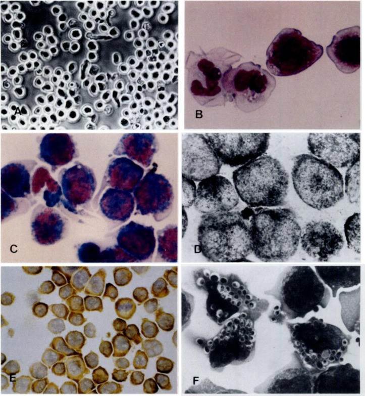 Fig. 1. (A) Inverted microscopic observation of PL-21 cells growing in single-cell suspension culture. (B) Smear of PL-21 cells stained with May-Grünwald-Giemsa (MGG). (C) Peroxidase staining of PL-21 cells. (D) Sudan Black B staining of PL-21 cells. (E) Lysozyme staining of PL-21 cells. (F) Phagocytosis of Candida albicans by PL-21 cells (Kubonishi I. et al., 1984).
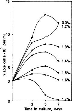 Fig. 2. Growth of PL-21 cells in various concentrations of DMSO. Viability was determined by trypan blue dye exclusions (Kubonishi I. et al., 1984).
Table 1. Differentiation of PL-21 cells in culture with or without 1.5% DMSO (Kubonishi I. et al., 1984).
Mybl, myeloblasts; Pro, promyelocytes; My, myelocytes; Met, metamyelocytes; Bnd, bands; Seg, segmented neutrophils.
| Duration of culture | Concentration of DMSO (%) | Differentiation (%) | Phagocytosis (%) | |||||
| Mybl | Pro | My | Met | Bnd | Seg | |||
| Day 0 | 0 | 2 | 96 | 1 | 0 | 0 | 1 | 6 |
| Day 5 | 0 | 0 | 83 | 9 | 4 | 3 | 1 | 11 |
| 1.5 | 0 | 54 | 12 | 19 | 10 | 5 | 51 | |
PTC596 Combination Treatment in AML Cell Lines
The prognosis for acute myeloid leukemia (AML) patients is poor, particularly in TP53 mutated AML, secondary, relapsed, and refractory AML, and in patients unfit for intensive treatment, thus highlighting an unmet need for novel therapeutic approaches. The combined use of compounds targeting the stem cell oncoprotein BMI1 and activating the tumor suppressor protein p53 may represent a promising novel treatment option for poor-risk AML patients.
Cell viability was determined in AML cell lines treated with increasing dosages of single compounds and in combination treatments using the BMI-1 inhibitor PTC596 and a variety of targeted therapies including the TP53 activator APR-246 (Fig. 3A), the MCL1 inhibitor S63845 (Fig. 3B), and the MEK inhibitor trametinib (Fig. 3C). AML cell lines represented all major morphologic and molecular subtypes including FLT3-ITD and FLT3 wild type, NPM1 mutant, and wild type, as well as TP53 mutant and wild type AML cell lines and a variety of patient-derived AML cells.
The combination of PTC596 and S63845 may be an effective treatment in CD34+ adverse risk AML with elevated MN1 gene expression and MCL1 protein levels, while PTC596 and trametinib may be more effective in CD34+ adverse risk AML with elevated BMI1 gene expression and MEK protein levels.
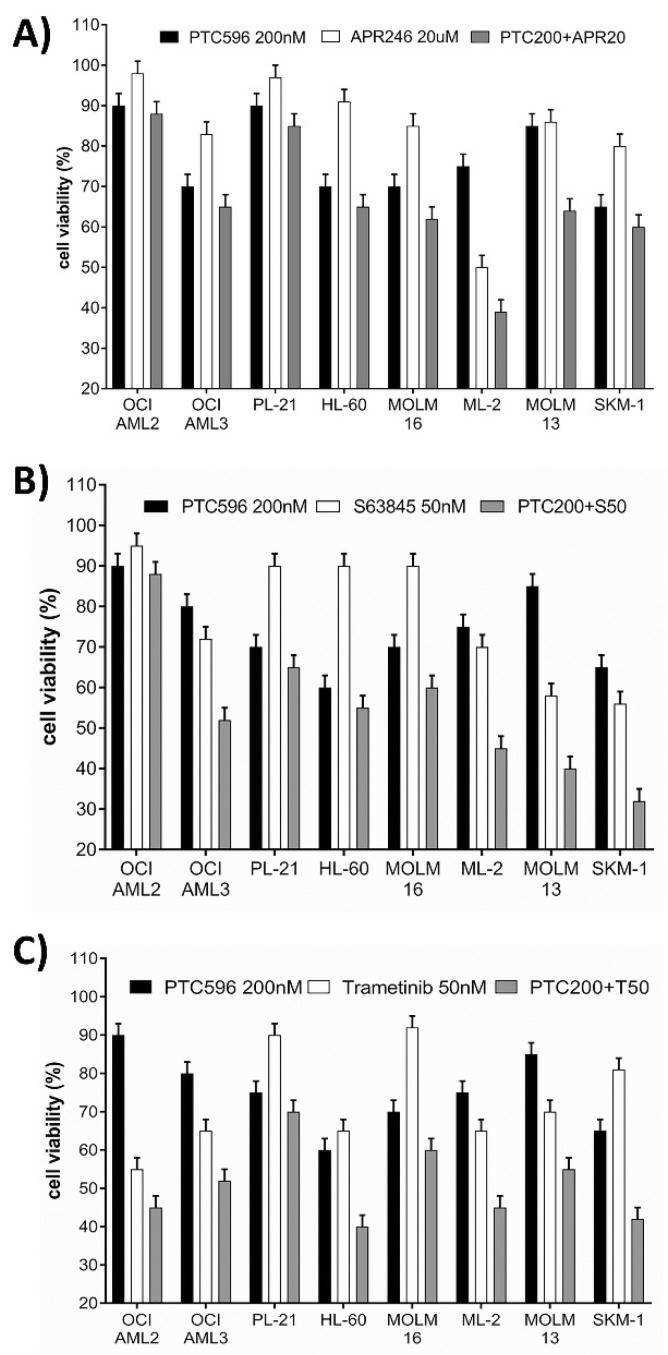 Fig. 3. Susceptibility of AML cell lines to various treatment combinations. Cell viability was determined in AML cells after 20 h treatment with single compounds and in combination with PTC596 and APR246 (A), PTC596 and S63845 (B), and PTC596 and trametinib (C) (Katja Seipel, et al., 2021).
Ask a Question
Write your own review
- You May Also Need

Description: Established in 2007 from the bone marrow mononuclear cells of an 82-year-old Japanese man with diffuse large B-cell lymphoma in the leukemic phase

Description: Established from the bone marrow of a 28-year-old man who developed the terminal leukemic phase of lymphosarcoma in 1976

Description: This cell line was derived from the bone marrow aspirate of a 59 year old male with erythroleukemia that became acute myelogenous leukaemia.The cells form colonies in soft-agar in the presence of ...

Description: Established from the pleural effusion of a 24-year-old woman with recurrent anaplastic large cell lymphoma (ALCL); cells were described to clonally derive from T-lineage lymphoid cells and to be ...

Description: Established from a 37-year-old man at second (refractory/terminal) relapse of Hodgkin lymphoma (nodular sclerosing -> lymphocyte depleted/stage IIISA -> stage IV) after both combined chemo- and ...

Description: Established from the peripheral blood of a 10-year-old Caucasian boy with acute lymphoblastic leukemia (pre B-ALL) at diagnosis in 1993
- Adipose Tissue-Derived Stem Cells
- Human Neurons
- Mouse Probe
- Whole Chromosome Painting Probes
- Hepatic Cells
- Renal Cells
- In Vitro ADME Kits
- Tissue Microarray
- Tissue Blocks
- Tissue Sections
- FFPE Cell Pellet
- Probe
- Centromere Probes
- Telomere Probes
- Satellite Enumeration Probes
- Subtelomere Specific Probes
- Bacterial Probes
- ISH/FISH Probes
- Exosome Isolation Kit
- Human Adult Stem Cells
- Mouse Stem Cells
- iPSCs
- Mouse Embryonic Stem Cells
- iPSC Differentiation Kits
- Mesenchymal Stem Cells
- Immortalized Human Cells
- Immortalized Murine Cells
- Cell Immortalization Kit
- Adipose Cells
- Cardiac Cells
- Dermal Cells
- Epidermal Cells
- Peripheral Blood Mononuclear Cells
- Umbilical Cord Cells
- Monkey Primary Cells
- Mouse Primary Cells
- Breast Tumor Cells
- Colorectal Tumor Cells
- Esophageal Tumor Cells
- Lung Tumor Cells
- Leukemia/Lymphoma/Myeloma Cells
- Ovarian Tumor Cells
- Pancreatic Tumor Cells
- Mouse Tumor Cells