Cell Proliferation Assay Services
- Service Details
- Features
- Applications
- FAQ
- Explore Other Options
Cell proliferation refers to the process of cell division and reproduction, which is the foundation for biological growth, development, reproduction, and heredity. It holds significant importance in fields such as cell biology, oncology, and pharmacology. Accurately measuring cell proliferation helps researchers understand cell growth characteristics, the effects of drugs on cell proliferation, and changes in the cell cycle.
With more than a decade of expertise in working with tumor cell lines, Creative Bioarray offers comprehensive cell proliferation assay services. We provide a range of assay methods tailored to specific cell types, protocols, and customer preferences for accurate proliferation measurement.

Our Cell Proliferation Assay Services
Workflow
Step 1: Requirement Analysis
Discuss experimental design based on customer needs, including cell type, detection methods, and time points.
Step 2: Experimental Design
Craft the optimal detection plan according to customer requirements.
Step 3: Sample Preparation and Handling
Prepare and handle samples following standard operating procedures.

Step 4: Experiment Execution
Perform experimental operations using appropriate cell proliferation detection methods.
Step 5: Data Analysis and Reporting
Fit proliferation curves, calculate IC50/EC50, conduct statistical significance analyses.
Methods
DNA Synthesis Detection
BrdU incorporation assay:
- Principle: Bromodeoxyuridine (BrdU), a thymidine analog, is incorporated into newly synthesized DNA strands during the S phase. Immunofluorescence and flow cytometry enable detection of BrdU-labeled cells which measure proliferation activity.
- Procedure: Cell culture → BrdU pulse labeling → Fixation/permeabilization → Anti-BrdU antibody incubation → Fluorescence detection/flow analysis.
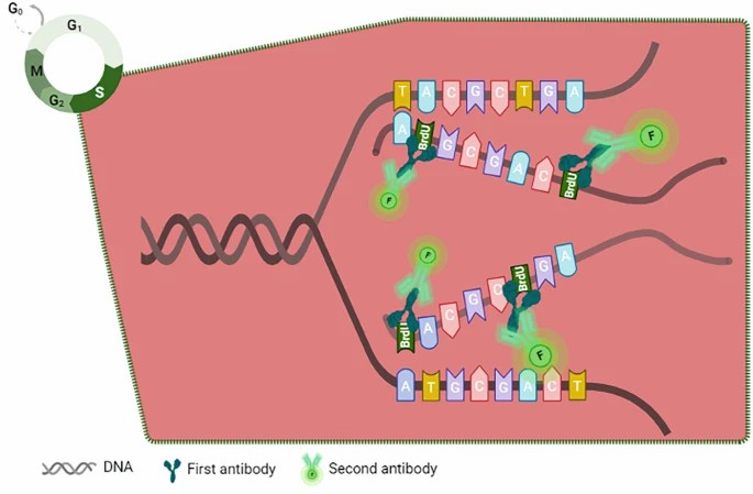 Fig. 1. The BrdU incorporation process (Yu JH, Wang ZX, et al., 2022).
EdU incorporation assay:
- Principle: 5-ethynyl-2'-deoxyuridine (EdU) is conjugated with fluorescent dyes via Click Chemistry to directly label newly synthesized DNA, offering higher sensitivity than BrdU for live or fixed cell detection.
- Procedure: Cell culture → EdU pulse labeling → Click reaction → Fluorescence microscopy imaging/flow analysis.
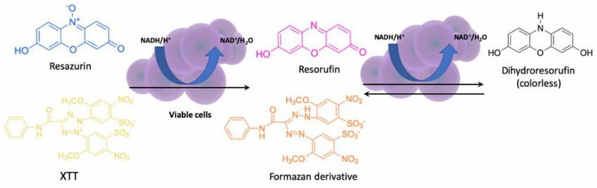 Fig. 2. The principle of XTT and resazurin assay (Jaśkiewicz M, Janczura A, et al., 2019).
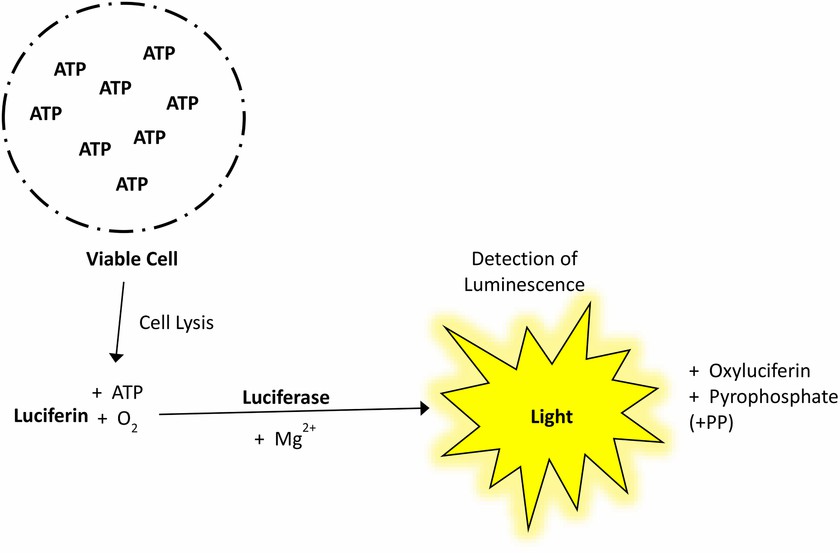 Fig. 3. Schematic illustration of the principles of ATP assay (Kamiloglu S, Sari G, et al., 2020).
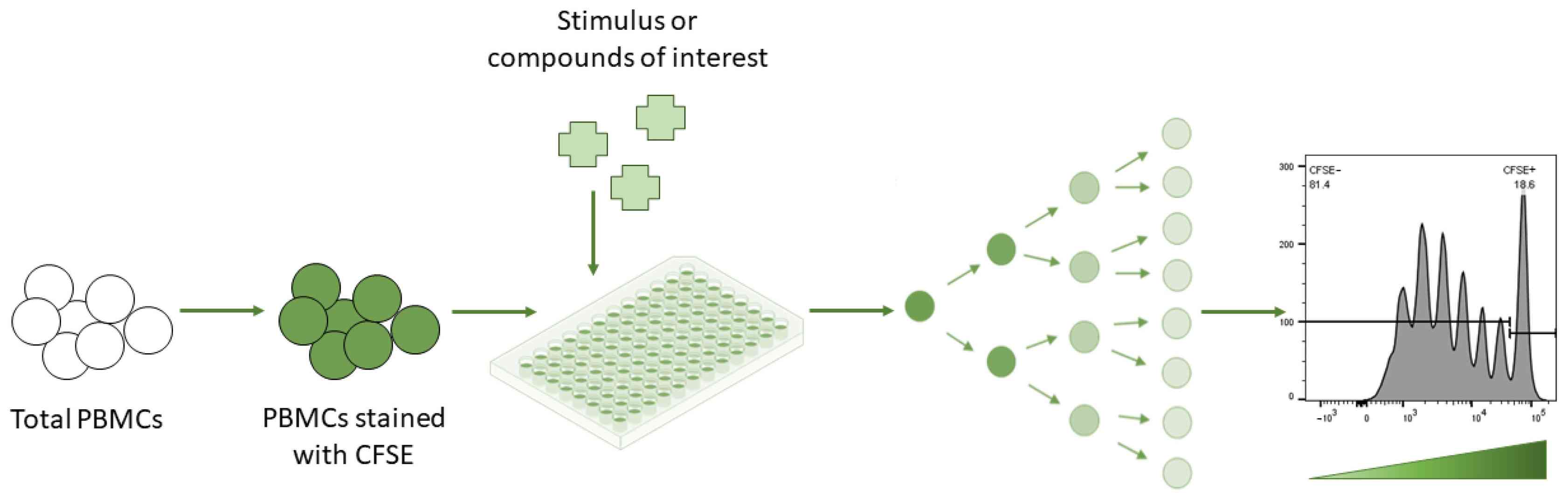 Fig. 4. Lymphocyte proliferation assay—CFSE assay (Ganesan N, Ronsmans S, et al., 2023)
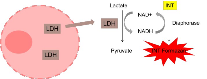 Fig. 5. Schematic representation of the principle of the LDH release assay (Forest V, Figarol A, et al., 2015).
Which Cell Proliferation Assay Should You Choose?
| Research Goal | Recommended Assay | Why This Works |
|---|---|---|
| High-throughput drug screening | ATP / MTT / XTT | Fast, scalable, quantitative |
| High sensitivity detection | EdU / ATP | Detects low cell numbers |
| Live-cell monitoring | Resazurin / Alamar Blue | Non-toxic, repeatable readouts |
| Cell cycle analysis | BrdU / EdU | Direct S-phase labeling |
| Immune cell proliferation | CFSE | Tracks division generations |
| Cytotoxicity confirmation | LDH + MTT | Distinguishes death vs growth inhibition |
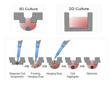
| Tumorigenicity studies | 3D Soft Agar | Mimics in vivo-like conditions |
| Mechanism/pathway validation | Ki-67 / PCNA | Links proliferation to signaling |
Creative Bioarray Cell Proliferation Assay Advantages

Flexible
Suitable assay methods chosen based on customer's cell type and requirements.

Accurate
Data strongly correlates to cell number.

Sensitive
Capable of detecting low cell numbers.

Fast
Enables high-throughput with quick results.
What can You do with Our Cell Proliferation Assay Services?
- Cancer Research: Evaluate drug efficacy in inhibiting tumor cell growth and aid in developing cancer therapies.
- Drug Development: Screen drug candidates for their impact on cell growth, ensuring safety and effectiveness.
- Toxicology Testing: Assess the effects of chemicals on cell proliferation to determine toxicity.
- Regenerative Medicine: Analyze stem cell growth for tissue engineering and therapy optimization.
- Basic Research: Study cellular mechanisms, such as cell cycle and signal transduction.
FAQ
1. How sensitive is cell proliferation detection?
The sensitivity varies with different detection methods. For example, fluorescence-based methods typically have higher sensitivity and can detect lower cell counts. We select the appropriate detection method based on the client's sample specifics before the experiment to ensure accurate results.
2. How are the results of cell proliferation detection presented?
We provide clients with a detailed experimental report, which includes the experimental methods, results (such as cell proliferation rate and cell count), data analysis, and conclusions. The results are typically presented in both charts and text format for easy understanding and use by clients.
3. Can I use specific cell lines?
Of course, we support the detection of various cell lines. You just need to provide detailed information when submitting your request.
Quotations and ordering
Our customer service representatives are available 24hr a day! We thank you for choosing Creative Bioarray service.
References
- Yu J, Wang Z, et al.BrdU Incorporation Assay to Analyze the Entry into S Phase. Methods Mol Biol. 2022. 2579:209-226.
- Forest V, Figarol A, et al. Adsorption of lactate dehydrogenase enzyme on carbon nanotubes: how to get accurate results for the cytotoxicity of these nanomaterials. Langmuir. 2015. 31(12):3635-43.
- Jaśkiewicz M, Janczura A, et al. Methods Used for the Eradication of Staphylococcal Biofilms. Antibiotics (Basel). 2019. 8(4):174.
- Kamiloglu S, Sari G, et al. Guidelines for cell viability assays. Food Frontiers. 2020.
- Ganesan N, Ronsmans S, et al. Methods to Assess Proliferation of Stimulated Human Lymphocytes In Vitro: A Narrative Review. Cells. 2023. 12(3):386.
Explore Other Options