In Vitro Cytotoxicity
- Service Details
- Workflow
- Features
- Study Example
- FAQ
- Explore Other Options
In vitro cytotoxicity testing using cultured cells is an essential tool for the early-stage safety assessment of pharmaceuticals, chemicals, biologics, and medical devices. An in vitro cytotoxicity assay enables the assessment of cell viability together with membrane integrity, cellular metabolic functions, and cell proliferation inhibition following test article exposure. Relative to in vivo assays, in vitro models are a rapid, cost-effective, and ethically sound method for initial toxicity screening, mechanism of action determination and selection of candidate compounds.
By employing cells and cell-based models derived from different tissues and organs, cytotoxicity testing can provide valuable insights into tissue-specific toxicity at an early stage of development. This approach allows high-throughput evaluation of candidate compounds, facilitating the identification of potential liabilities and enabling the prioritization of safer and more promising molecules.
All of our cytotoxicity assays are conducted in accordance with international regulatory guidelines and are designed to help you make informed, data-driven decisions throughout your R&D process.
Our In Vitro Cytotoxicity Service Content:
We offer a comprehensive suite of in vitro cytotoxicity assays, covering multiple toxicological endpoints:
Cell Viability Assays: MTT, WST-1, CCK-8, Resazurin-based (Alamar Blue), Crystal violet staining' etc.
Membrane Integrity Assays: LDH release assay, Trypan Blue exclusion
Apoptosis/Necrosis Detection: Annexin V/PI staining, TUNEL assay, Caspase 3/7 activity
Mitochondrial Function Assays: JC-1 dye for mitochondrial membrane potential, ATP quantification, MitoTracker staining
High-Content Imaging: Multiparametric analysis of cell morphology, membrane damage, and nuclear condensation
Available models
- Standard Cell Lines: Such as HepG2, SH-SY5Y, SK-N-SH, PC-3, HK2, NHBE, HaCaT, L-929, HEK293, A549.
- Primary Cell Lines: Such as primary hepatocytes, renal epithelial cells, cardiomyocytes, hematopoietic cells, PBMCs, bone marrow cells.
- 3D Culture Systems & Organoids: Brain, heart, lung, liver, kidney, and intestinal organoids. Blood vessel, retina. Tumor spheroids for enhanced physiological relevance.
- Microfluidic Cell Culture Systems: Dynamic organ-on-a-chip models for perfusion-based toxicity studies
Workflow

Step 1: Consultation & Project Design
Discuss compound type, exposure route, regulatory goals, and study design.

Step 2: Cell Line Selection & Assay Setup
Choose appropriate cell models and validate assay protocols.

Step 3: Dose-Response Testing
Test multiple concentrations to determine IC₅₀/EC₅₀ or cytotoxicity thresholds.


Step 4: Data Analysis
Statistical analysis, dose-response curve fitting, endpoint comparison.

Step 5: Report Delivery
Detailed study report with raw data, experimental protocol, result interpretation, and representative images.
Features

Flexible & Customizable
Tailored assay design based on compound class, exposure route, and regulatory goals

Experienced Team
Our toxicology scientists are experts in cell-based assay development and data interpretation.

High-Quality Data Output
Validated assays, positive/negative controls and rigorous reproduction to ensure accurate and reliable results.

Fast Turnaround
Optimized workflows and real-time updates to speed up your research timeline.
Study Example
Small molecule activation of m6A mRNA methylation as a novel approach for neuroprotection
Background
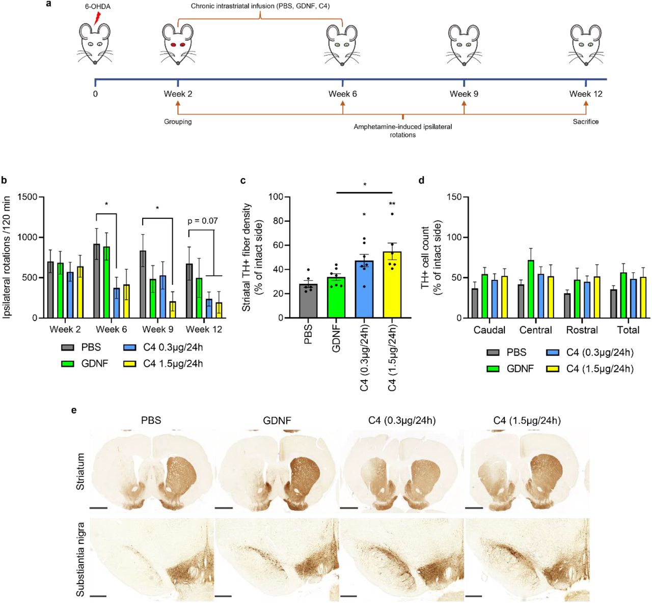
This bioRxiv preprint investigates small-molecule activation of m6A mRNA methylation for neuroprotection. The authors previously identified METTL3/14/WTAP activators. Here, C3 and C4 supported survival of mouse dopamine neurons at nanomolar concentrations, while METTL3/14 inhibitor STM2457 caused death. C4 protected human iPSC-derived dopamine neurons from 6-OHDA, enhanced neurite outgrowth, and improved motor behavior in the rat 6-OHDA Parkinson's disease model more efficiently than GDNF. This first demonstrates that m6A regulators can protect and regenerate neurons, suggesting a novel therapeutic pathway for Parkinson's disease.
Key Findings
METTL3/14 activators C3 and C4 protected mouse dopamine neurons from growth factor deprivation and 6-OHDA-induced death at 10 nM. In contrast, the METTL3/14 inhibitor STM2457 triggered dopamine neuron death. C4 protected human iPSC-derived dopamine neurons from 6-OHDA and increased neurite outgrowth and process number. In the rat 6-OHDA Parkinson's disease model, C4 improved motor behavior and protected dopamine neurons and fibers faster and more efficiently than GDNF. Molecular dynamics showed C4 stabilizes SAM binding to METTL3/14, especially a Tyr406 π-cation interaction. C4 failed to protect peripheral SCG and DRG neurons when applied extracellularly, but microinjection rescued them, suggesting limited membrane penetration. These are the first specific METTL3/14 activators and the first demonstration that m6A regulators can protect and regenerate neurons.
Contribution
Creative Bioarray performed the cytotoxicity screening of compound C4 (Fig. 1c), assessing endpoints including cell count, nuclear size, DNA structure, cell membrane permeability, mitochondrial mass, mitochondrial membrane potential, and cytochrome c release. Results showed no aberrations up to 200 μM, supporting C4's safety profile.
FAQ
Which samples can be tested in your in vitro cytotoxicity assays?
Small molecules, peptides, biologics, nanoparticles, medical device extracts, and other chemical formulations can all be tested.
What kind of sample should I provide for testing?
Please provide pure samples of your compound (either powder or solution) along with basic physicochemical information, including solubility, to ensure optimal testing conditions.
Do you provide time-dependent toxicity (TDI) testing?
Yes, we can collect data at multiple time points (4 h, 24 h, 48 h, 72 h) to assess acute versus delayed toxicity and calculate AUC-based IC₅₀ values to distinguish the two.
Do you provide high-throughput screening (HTS) capability?
Yes, our fully automated 384-well workstation has the capability for high-throughput testing and can process over 2000 wells per day, making us well suited to assist with compound library screening, QC, and STR studies.
Explore Other Options