Brain Organoid Differentiation Service from iPSC
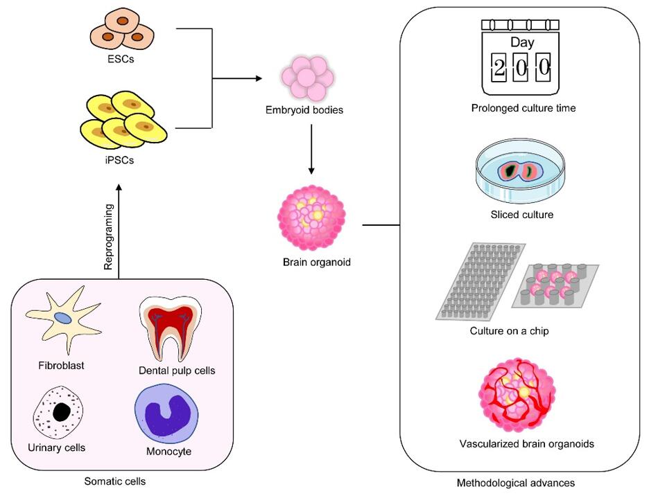
While induced pluripotent stem cells (iPSCs) have replaced the ethically questionable human embryonic stem cells, iPSC-based neuronal differentiation studies remain descriptive at the cellular level but fail to adequately provide the details that could be derived from a complex, 3D human brain tissue. Brain organoids have emerged as an excellent model for investigating brain development and the mechanisms of related diseases.
Creative Bioarray developed a protocol that is able to differentiate brain organoids from induced Pluripotent Stem Cells(iPSCs). We also provide characterization of iPSC-derived organoids using immunocytochemistry and multi-electrode array (MEA).
iPSC-derived brain organoids can be used for
- Exploring mechanisms for neurological disorders
- Drug screening
- Modeling brain disorders
- Development of organ-chips
We provide various types of brain organoids:
- Microglia-enriched brain organoid
- Oligodendrocytes brain organoid
- Blood-vessel-enriched brain organoid
The brain organoids we deliver are a mixture of various neuronal cells, and the mixing ratio can be adjusted according to customer requirements.
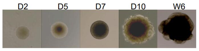 Figure 1.
Phase contrast images taken after differentiation, showing the morphological changes occurred.
Figure 1.
Phase contrast images taken after differentiation, showing the morphological changes occurred.
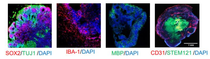 Figure 2. Differentiation of different types of
brain organoids.
Figure 2. Differentiation of different types of
brain organoids.
Creative Bioarray also provides iPSC-derived brain organoids as ready-to-use products, as well as the specialized medium to support the optimal performance of the organoids. If you have any need for Brain Organoid Differentiation service, do not hesitate to contact us for this special service. Please let us know what you need and we will accommodate you. We look forward to working with you.
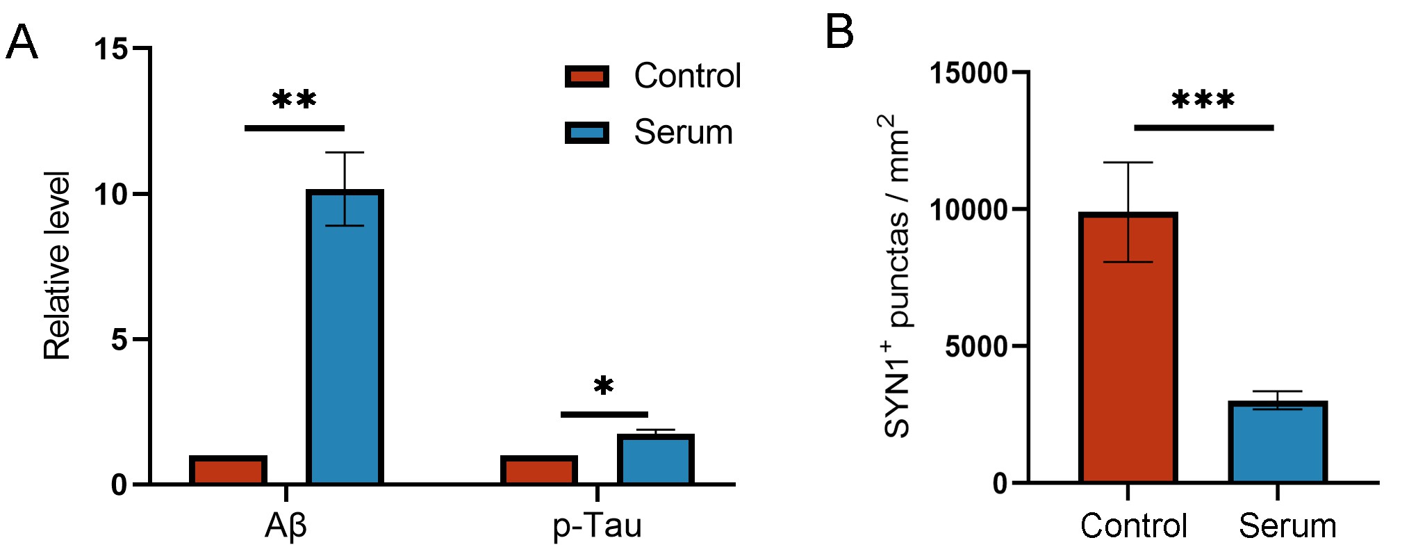
Case Studies
Case 1: iPSC-derived brain organoids exposed to serum exhibited increased levels of amyloid-beta (Aβ) aggregates and phosphorylated microtubule‐associated tau protein (p‐Tau) (A). The high-content cellular analysis platform demonstrated a significant reduction in synapses (B), indicating the formation of Alzheimer's disease (AD)-like pathology, which can be utilized for constructing sporadic AD models.
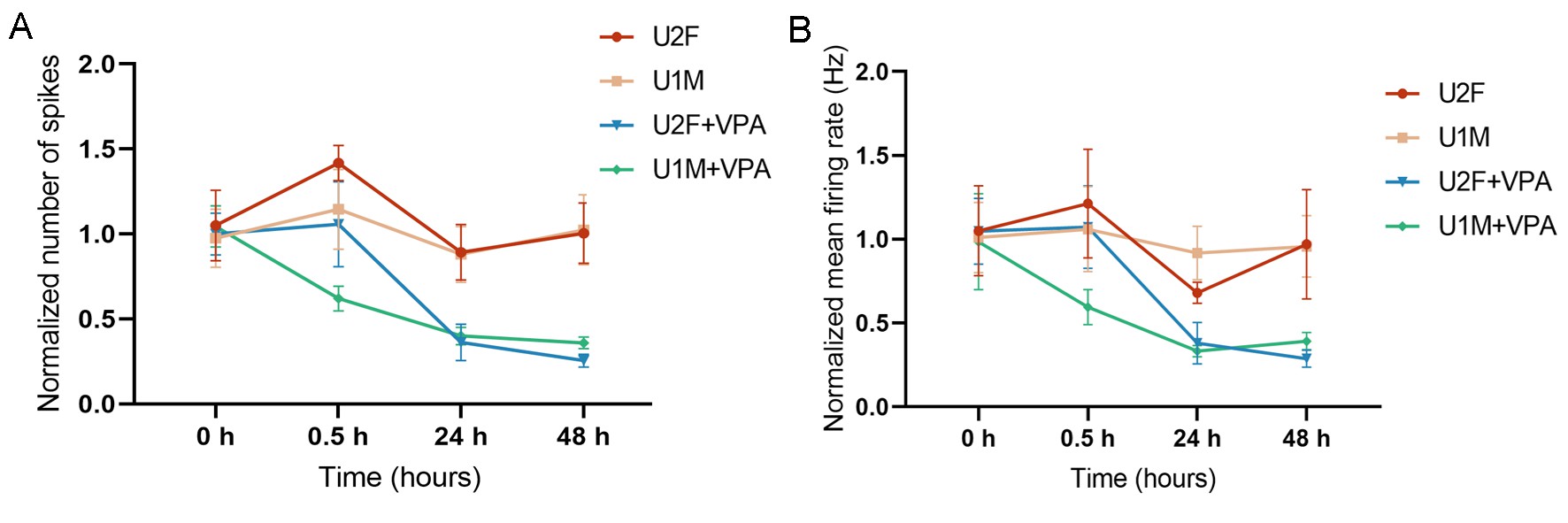
Case 2: Using our high-throughput electrophysiological platform to analyze brain organoids exposed to Valproic Acid (VPA), there was a significant decrease in the number of spikes and mean firing rate, which is of significant importance for studying the pathogenesis of ASD.
Explore Other Options