Noise-Induced Hearing Loss (NIHL) Model
Creative Bioarray has established a robust and reliable Noise-Induced Hearing Loss (NIHL) Model that is transforming auditory research. This advanced model is an essential tool for researchers delving into the complexities of hearing impairment and for testing the efficacy of therapeutic compounds. Unlock unprecedented insights into auditory health with our cutting-edge NIHL Model, which serves as a foundational element for breakthroughs in the diagnosis and treatment of auditory disorders. Partner with us to elevate your research capabilities.
What is Noise-Induced Hearing Loss (NIHL)?
NIHL is the second most prevalent cause of sensorineural hearing loss, following age-related hearing loss, and impacts approximately 5% of the global population. NIHL carries significant physical, mental, social, and economic consequences for both individuals and society. Factors such as stress and social isolation in patients' workplaces and personal lives can lead to declines in quality of life that often go unnoticed. The pathophysiology of NIHL is complex and multifactorial, involving both genetic and environmental influences, with considerable contributions from occupational exposure. Current research into pharmacological treatments for NIHL explores various agents, including anti-inflammatory, antioxidant, anti-excitatory, and anti-apoptotic compounds. Despite notable progress in understanding the underlying mechanisms of NIHL, there is still limited evidence supporting effective pharmacotherapeutic interventions.
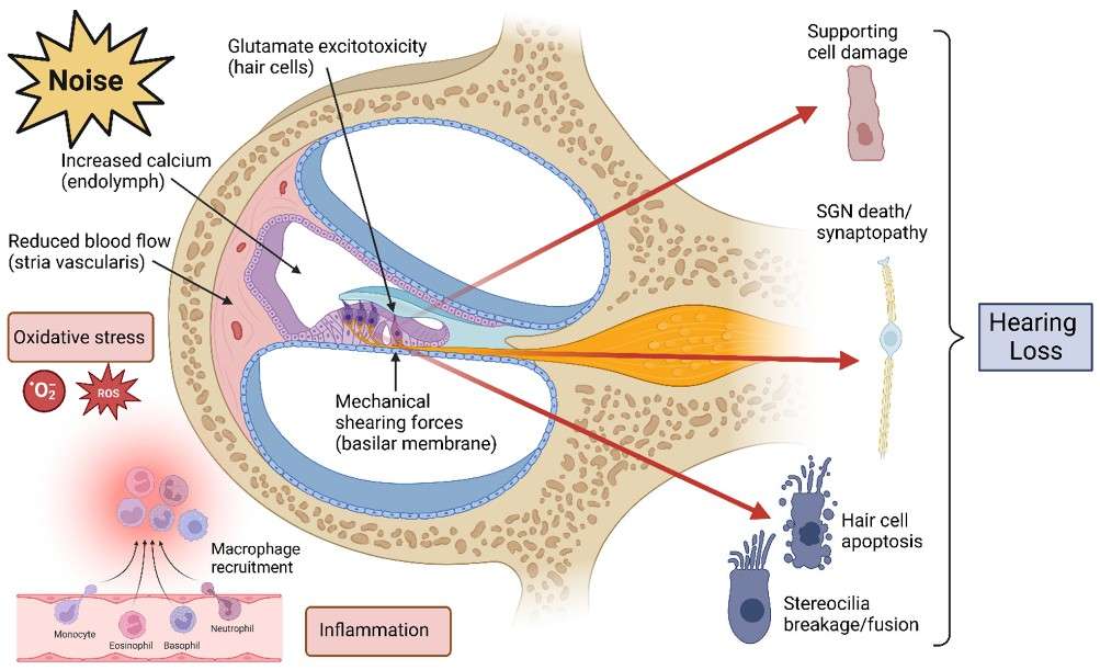 Fig. 1: Mechanisms of damage in NIHL. (Natarajan et al., 2023)
Creative Bioarray's Noise-Induced Hearing Loss (NIHL) Model
Available Animal
- Rat
- Mouse
Modeling Method
-
All animals undergo noise exposure to establish the hearing loss model.
Endpoints
- Auditory brainstem response (ABR) measurement
- Distortion product otoacoustic emission (DPOAE)
- Histology analysis
- qPCR or Western blot
- Customized endpoints tailored to your specific research requirements
Example Data
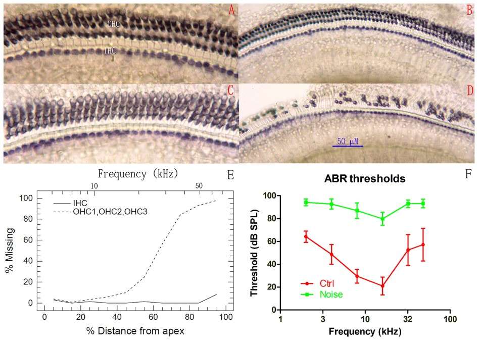 Fig. 2 Noise-induced hair cell loss and ABR threshold shift measured 3 months after the noise exposure at 6–8 weeks of age. (A-D) Representative images of SDH staining of HCs from the two groups. (A, B) Apical and basal turns, respectively, in the control group; (C, D) Apical and basal turns, respectively, in the noise group. Massive OHC loss is clearly observed in (D). (E) Cochleogram of the noise-exposed animals showing the loss of OHCs concentrated in the basal half of the cochlea. (F) ABR thresholds. (Liu et al. 2016)
Quotation and Ordering
Creative Bioarray is making significant strides in advancing the field of auditory research by providing an extensive selection of validated in vivo models. If you are interested in our services, please feel free to contact us at any time or submit an inquiry to us directly. Our team is ready to assist you in navigating your research needs.
References
- Natarajan, N., et al. Noise-induced hearing loss. Journal of clinical medicine, 2023, 12(6): 2347.
- Liu, L., et al. Noise induced hearing loss impairs spatial learning/memory and hippocampal neurogenesis in mice. Scientific reports, 2016, 6(1): 20374.