Cell Viability Assays
- Service details
- Features
- FAQ
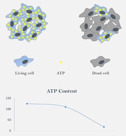
Cell viability assays are essential for evaluating cellular health, metabolic activity, and responsiveness to external stimuli. These assays are widely applied in drug development, toxicity testing, cancer research, and regenerative medicine. Cell viability is quantified by measuring the proportion of living versus dead cells within a population. Increased viability typically correlates with cell proliferation, while reduced viability may indicate compound toxicity or suboptimal culture conditions.
By assessing metabolic capacity, membrane integrity, enzymatic activity, and ATP levels, researchers gain precise insights into cellular health and survival. At Creative Bioarray, we deliver comprehensive, efficient, and accurate cell viability testing solutions tailored to diverse research needs.
Services
Our cell viability assays encompass both classical and advanced methodologies to address varied experimental requirements:
Tetrazolium Reduction Assays (MTT/MTS/WST-8/WST-1/CCK-8)
These assays measure cellular metabolic activity through the reduction of tetrazolium salts to colored formazan products by mitochondrial dehydrogenases in viable cells. Quantification is performed via spectrophotometric or microplate reader absorbance measurements.
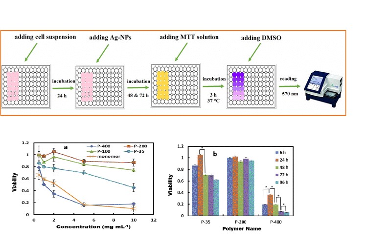 Fig. 1. Schematic diagram for MTT assay of synthesized Ag-NPs by P. farcta extract at
different incubation times (Miri A and Sarani M, 2018).
Fig. 1. Schematic diagram for MTT assay of synthesized Ag-NPs by P. farcta extract at
different incubation times (Miri A and Sarani M, 2018).
Membrane Integrity Assays
Fluorescent dyes (e.g., propidium iodide, PI) or trypan blue staining distinguish live/dead cells based on membrane integrity. Viable cells exclude dyes, while compromised membranes in dead cells permit dye uptake.
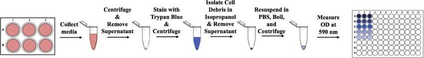 Fig. 2. Trypan blue cellular debris assay (Lebeau PF, Chen J, et al., 2019).
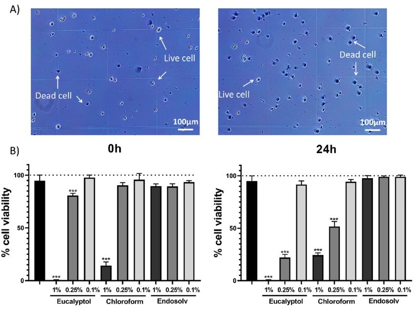 Fig. 3. Trypan blue assay results (Sanz JL, López-García S, et al., 2022).
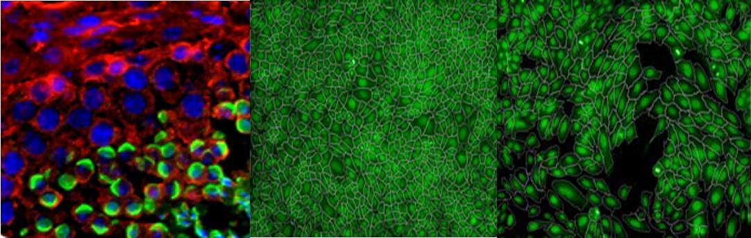
Enzymatic Activity Substrates
Fluorogenic or chromogenic substrates detect specific intracellular enzyme activities. For example, non-fluorescent Calcein-AM is converted to fluorescent calcein by esterases in live cells.
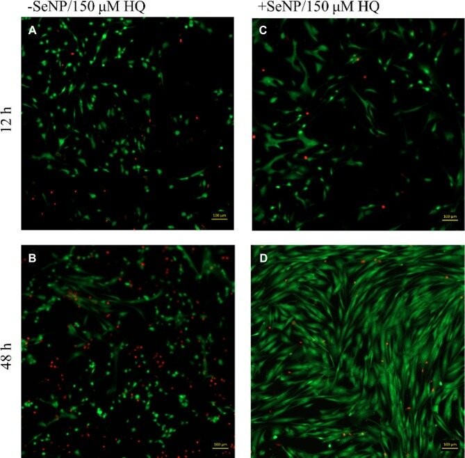 Fig. 4. Live/dead images (calcein AM/ethidium homodimer-1) images for human dermal
fibroblasts (HDFs) with 150 μM HQ challenge at 12 and 48 hrs (Chung S, Roy AK,
et al., 2019).
Fig. 4. Live/dead images (calcein AM/ethidium homodimer-1) images for human dermal
fibroblasts (HDFs) with 150 μM HQ challenge at 12 and 48 hrs (Chung S, Roy AK,
et al., 2019).
ATP Determination
Utilizing the luciferase-luciferin system, this method quantifies intracellular ATP levels with high sensitivity, ideal for low-metabolic cells or rapid viability assessments.
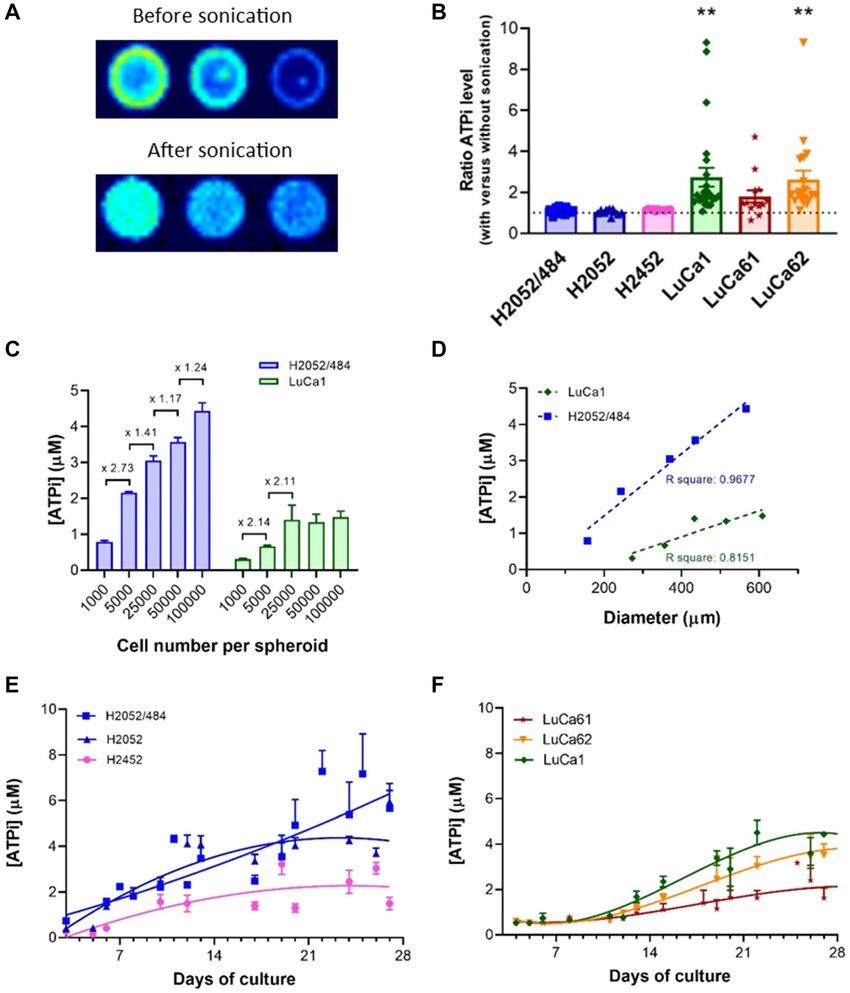 Fig. 5. Cell viability measurement using an ATP-based luminescence assay (Gendre DAJ,
Ameti E, et al., 2021).
Fig. 5. Cell viability measurement using an ATP-based luminescence assay (Gendre DAJ,
Ameti E, et al., 2021).
Single/Two Color Viability Assays
Dual-fluorescence labeling (e.g., AO/PI staining) enables simultaneous differentiation of live (acridine orange) and dead (propidium iodide) cells, providing detailed viability profiles.
Microplate Cell Viability Assays
High-throughput 96/384-well plate formats compatible with colorimetric (MTT, XTT, CCK-8) or fluorescent readouts, optimized for large-scale drug screening and proliferation studies.
Custom Solutions
Creative Bioarray designs tailored cell-based assays using UV/Vis, fluorescence, or luminescence detection to address your unique research goals.
Features

Comprehensive Solutions
Diverse methodologies adaptable to all cell types and experimental designs.

High Sensitivity & Accuracy
State-of-the-art instrumentation and premium-grade reagents ensure reproducibility.

Customized Services
Flexible experimental design and expert technical support aligned with your objectives.
FAQ
How do I select the optimal assay for my study?
Experimental objectives (such as metabolic activity or membrane integrity) and the specific cell type determine the optimal assay selection. MTT/WST-8 assays are efficient for high-throughput metabolic assessments and ATP assays work best in low-metabolic systems while dual-color staining allows for visual viability assessment. Our specialists will guide your selection.
What cell types are compatible with your assays?
Our services support mammalian cells (including tumor cells and stem cells), primary cultures, bacteria, and yeast. Reach out to us to obtain tailored solutions for your unique cell models.
How are results delivered?
Reports include detailed protocols, raw data (absorbance/fluorescence readings), analyzed results (viability percentages, dose-response curves), and expert interpretation. Data formats (e.g., GraphPad Prism files) are available upon request.
What causes low cell viability in results?
Potential factors include suboptimal culture conditions, cellular senescence, compound toxicity, or assay incompatibility. Our team provides root-cause analysis and optimization strategies to refine your workflow.
References
- Miri A, Sarani M. Biological studies of synthesized silver nanoparticles using Prosopis farcta. Mol Biol Rep. 2018. 45(6):1621-1626.
- Lebeau PF, Chen J, et al. The trypan blue cellular debris assay: a novel low-cost method for the rapid quantification of cell death. MethodsX. 2019. 6:1174-1180.
- Sanz JL, López-García S, et al. Are Endodontic Solvents Cytotoxic? An In Vitro Study on Human Periodontal Ligament Stem Cells. Pharmaceutics. 2022. 14(11):2415.
- Chung S, Roy AK, et al. Selenium Nanoparticle Protection of Fibroblast Stress: Activation of ATF4 and Bcl-xL Expression. Int J Nanomedicine. 2019. 14:9995-10007.
- Gendre DAJ, Ameti E, et al. Optimization of tumor spheroid model in mesothelioma and lung cancers and anti-cancer drug testing in H2052/484 spheroids. Oncotarget. 2021. 12(24):2375-2387.
Related Services
Explore Other Options