
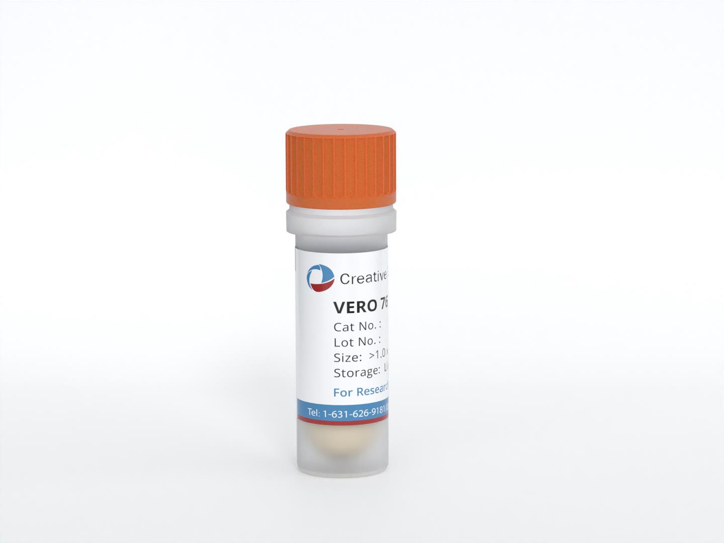
VERO 76
Cat.No.: CSC-C9750L
Species: Chlorocebus sabaeus (Green monkey)
Source: Kidney
Morphology: epithelial
Culture Properties: monolayer
- Specification
- Background
- Scientific Data
- Q & A
- Customer Review
Virus: several hemorrhagic fever viruses including; Machupo virus, Junin virus; Lassa fever virus; Marburg virus; Ebola virus
Histopathology: normal
VERO 76 is a well-characterized continuous cell line derived from the kidney of an African green monkey (Cercopithecus aethiops). Established in 1968 by K.M. Johnson, it is a derivative of the original Vero line.
A key distinguishing feature of VERO 76 is its lower saturation density compared to the parental Vero line, which can be advantageous for specific experimental designs. More critically, like other Vero sublines, VERO 76 harbors a large deletion and loss of heterozygosity on chromosome 12 encompassing the type I interferon gene cluster. This interferon deficiency prevents the cells from producing interferon-α or -β in response to viral infection, enabling robust viral replication and clear cytopathic effect (CPE) observation—making it an exceptionally sensitive system for antiviral compound screening.
VERO 76 exhibits broad susceptibility to a diverse range of viruses. It supports replication of human haemorrhagic fever viruses including Ebola, Marburg, Lassa, Junin, and Machupo viruses, producing visible plaques upon infection. The line has also been successfully employed in plaque assays for Rickettsia prowazekii and in neutralization assays for vaccine development. Its ease of culture in standard DMEM-based medium further facilitates routine laboratory use.
These attributes—interferon deficiency, broad viral susceptibility, well-characterized genetic background, and practical handling—establish VERO 76 as a versatile and reliable platform bridging fundamental virology, vaccine development, and antiviral drug discovery.
Evaluation of the Cytotoxicity of Antimicrobial Lipopeptides
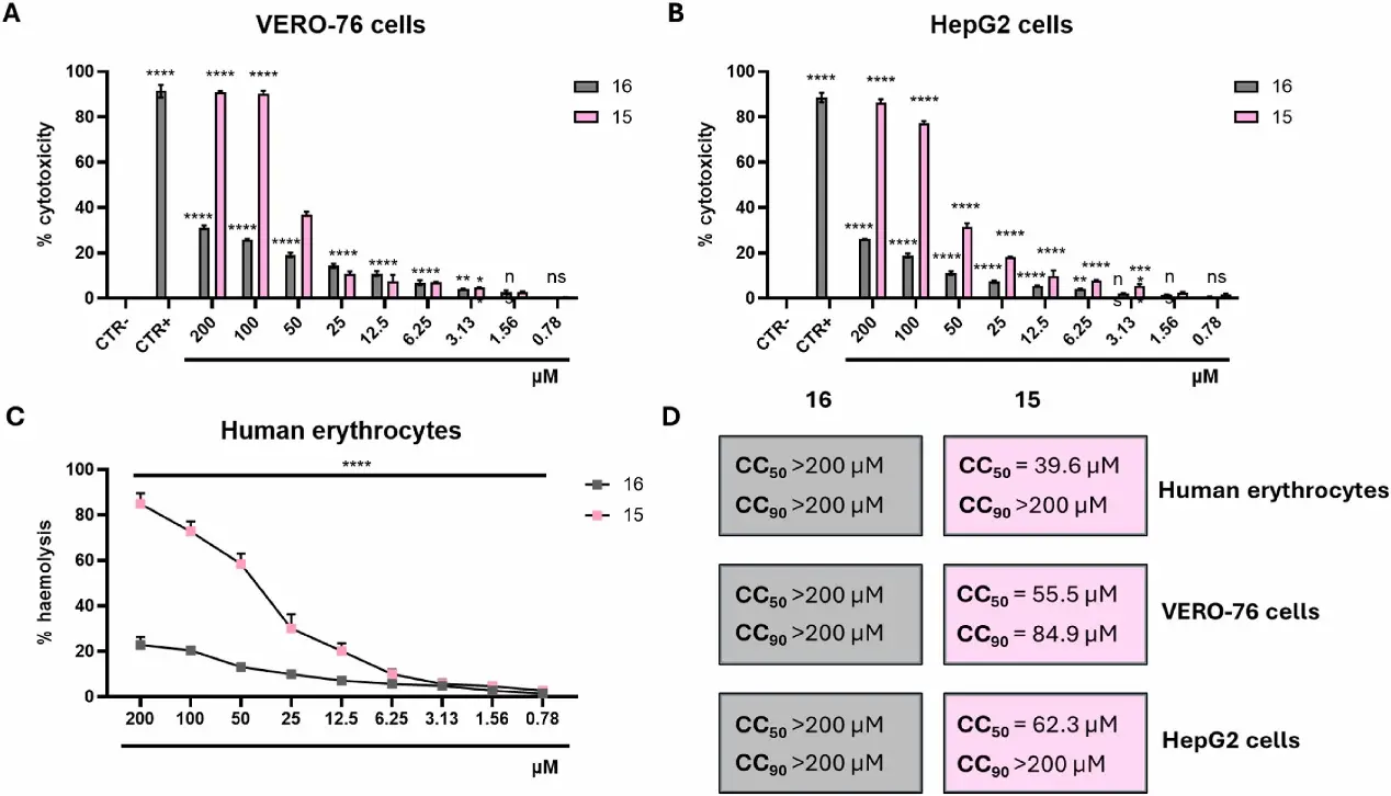
The most severe adverse effects of antimicrobial lipopeptides are generally related to their haemolytic activity and cytotoxicity, inherent to their antibacterial mechanism of action. To evaluate potential unintended, non-specific membrane lysis promoted by the most active compound 15, its toxicity was assessed using MTT and haemolysis assays on human renal (VERO-76), hepatic (HepG2), and erythrocyte cells across a concentration range of 0.78 to 200 μM. Compound 16, the inactive analogue, was used as a negative control (CTR–) while 0.1% Triton X-100 and 100% DMSO were employed as positive controls (CTR+). As expected, 16 induced comparatively weak cytotoxicity in VERO-76 (Fig. 1A) and HepG2 cells (Fig. 1B) at the maximum concentration, with cell mortality of 31.3% and 26.0%, respectively. In contrast, compound 15 exhibited higher toxicity compared to 16, causing 91.1% and 86.3% cell death in VERO-76 and HepG2 cells, respectively, at 200 μM. Despite this enhanced toxicity, its 50% cytotoxic concentration (CC50) was calculated to be 55.5 μM for VERO-76 cells and 84.9 μM for HepG2 cells, approximately 20 times higher than its MIC values, indicating a potentially favourable therapeutic window. Erythrocyte lysis followed a similar trend with 15 causing more pronounced lysis than 16. The CC50 value for compound 15 in the haemolysis assay was 39.6 μM (Fig. 1C), approximately 10-folds higher than its MIC, hence retaining an acceptable therapeutic range (Fig. 1D).
Ask a Question
Write your own review
- You May Also Need
Description: Species: monkey - African Green monkeyStrain: Cercopithecus aethiopsVirus Susceptibility: SV40; poliovirus 1; vesicular stomatitis (Indiana)Production: keratinHistopathology: normalNote: the cells ...
Description: Derived from a pooled cell suspension from kidneys removed from six adult Rhesus monkeys; the line is susceptible to a broad range of viruses that includes members of the enterovirus, rhinovirus, ...
Description: Established from an African Green monkey foetal kidney. The cells are highly susceptible to Simian rotavirus SA11
Description: Established from CV-1 Simian cells (cercopithecus aethiops) which were transformed by an origin-defective mutant of SV-40; cells are host cells for the propagation of pure populations of recombinant ...
Description: Established from peripheral blood lymphocytes of a cotton-top tamarin (cotton-top marmoset) monkey (Saguinus oedipus); cells were described to release high titers of Epstein-Barr virus (EBV) ...
- Adipose Tissue-Derived Stem Cells
- Human Neurons
- Mouse Probe
- Whole Chromosome Painting Probes
- Hepatic Cells
- Renal Cells
- In Vitro ADME Kits
- Tissue Microarray
- Tissue Blocks
- Tissue Sections
- FFPE Cell Pellet
- Probe
- Centromere Probes
- Telomere Probes
- Satellite Enumeration Probes
- Subtelomere Specific Probes
- Bacterial Probes
- ISH/FISH Probes
- Exosome Isolation Kit
- Human Adult Stem Cells
- Mouse Stem Cells
- iPSCs
- Mouse Embryonic Stem Cells
- iPSC Differentiation Kits
- Mesenchymal Stem Cells
- Immortalized Human Cells
- Immortalized Murine Cells
- Cell Immortalization Kit
- Adipose Cells
- Cardiac Cells
- Dermal Cells
- Epidermal Cells
- Peripheral Blood Mononuclear Cells
- Umbilical Cord Cells
- Monkey Primary Cells
- Mouse Primary Cells
- Breast Tumor Cells
- Colorectal Tumor Cells
- Esophageal Tumor Cells
- Lung Tumor Cells
- Leukemia/Lymphoma/Myeloma Cells
- Ovarian Tumor Cells
- Pancreatic Tumor Cells
- Mouse Tumor Cells