Exosome Quantification
In the past decade, exosomes have received much attention due to their potential applications in molecular biology, and biomedical fields. The accurate and reliable quantification of exosomes is crucial to better understand exosomes and their relationship with various diseases.
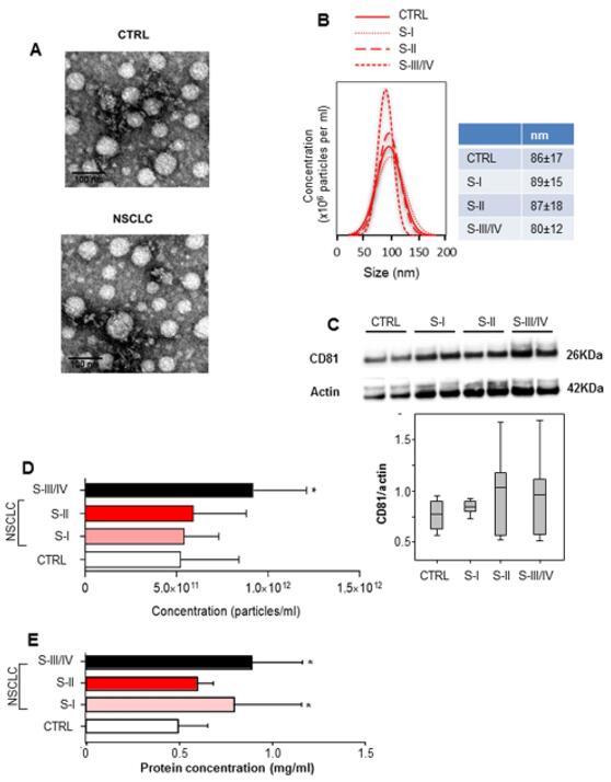 Figure 1 Characterization and quantifcation of exosome levels in serum samples from healthy controls and non-small cell lung cancer (NSCLC) patients.
Exosome quantification mainly depends on their characteristic physical properties, such as size, mass, and density, or on membrane proteins presented on their surface. Creative Bioarray offers a range of options to meet most exosome quantitation demands. We provide reliable, and optimized tools at the most competitive price for quantitative analysis of exosomes in diverse biological samples such as plasma, urine, etc.
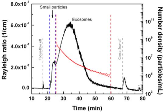 Figure 2 An AF4-MALS fractogram of exosome sample and calculated number of particles (number density of particles per mL) for larger exosome population as a function of elution time.
Here in Creative Bioarray, we provide a series of exosome quantification services listed as below, choose the exosome quantitation method that is best for your studies:
- ELISA-based Immunoaffinity capture (IAC) assay
- Nanoparticle tracking analysis (NTA)
- Asymmetrical-flow field flow fractionation (AF4) coupled with multidetection assay
- Dynamic light scattering (DLS) assay
- Surface plasmon resonance (SPR) assay
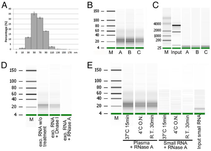 Figure 3 Exosome isolation and exosomal small RNA quantification.
In Creative Bioarray, we also provide exosomal RNA quantification service. If you have any special needs in our Exosome Quantification Service, please contact us. Let us know what you need and we will accommodate you. We look forward to working with you in the future.
References
- Grimolizzi, F.; et al. Exosomal miR-126 as a circulating biomarker in non-small-cell lung cancer regulating cancer progression. Sci Rep. 2017, 7(1): 15277.
- Sitar, S.; et al. Size characterization and quantification of exosomes by asymmetrical-flow field-flow fractionation. Anal Chem. 2015, 87(18): 9225-9233.
- Huang, X.; et al. Characterization of human plasma-derived exosomal RNAs by deep sequencing. BMC Genomics. 2013, 14(1): 319.
Explore Other Options