Hypoxic-Ischemic Encephalopathy (HIE) Model
- Background
- Models
- Study Examples
- Features
- FAQ
Service Overview
Creative Bioarray offers standardized Hypoxic-Ischemic Encephalopathy (HIE) animal model services based on the well-established Rice–Vannucci neonatal brain injury model. Our platform is optimized for preclinical neuroprotection, neuroinflammation, and CNS drug efficacy evaluation, providing high-quality and reproducible in vivo data for translational neuroscience research.
We support rat (P7–P10) and mouse models (C57BL/6, CD-1) to simulate neonatal hypoxic-ischemic brain injury with high clinical relevance. The model enables robust evaluation of therapeutic candidates targeting brain injury, inflammation, and functional recovery.
Overview of Hypoxic-Ischemic Encephalopathy (HIE)
Neonatal Hypoxic-Ischemic Encephalopathy remains a devastating cause of infant mortality and long-term neurological deficits like cerebral palsy. The condition is triggered by a sudden reduction in oxygen and blood flow to the brain, initiating a "double-hit" injury. An initial primary energy failure causes immediate cell death, followed by a secondary energy failure hours later, driven by excitotoxicity, oxidative stress, and a massive inflammatory cascade.
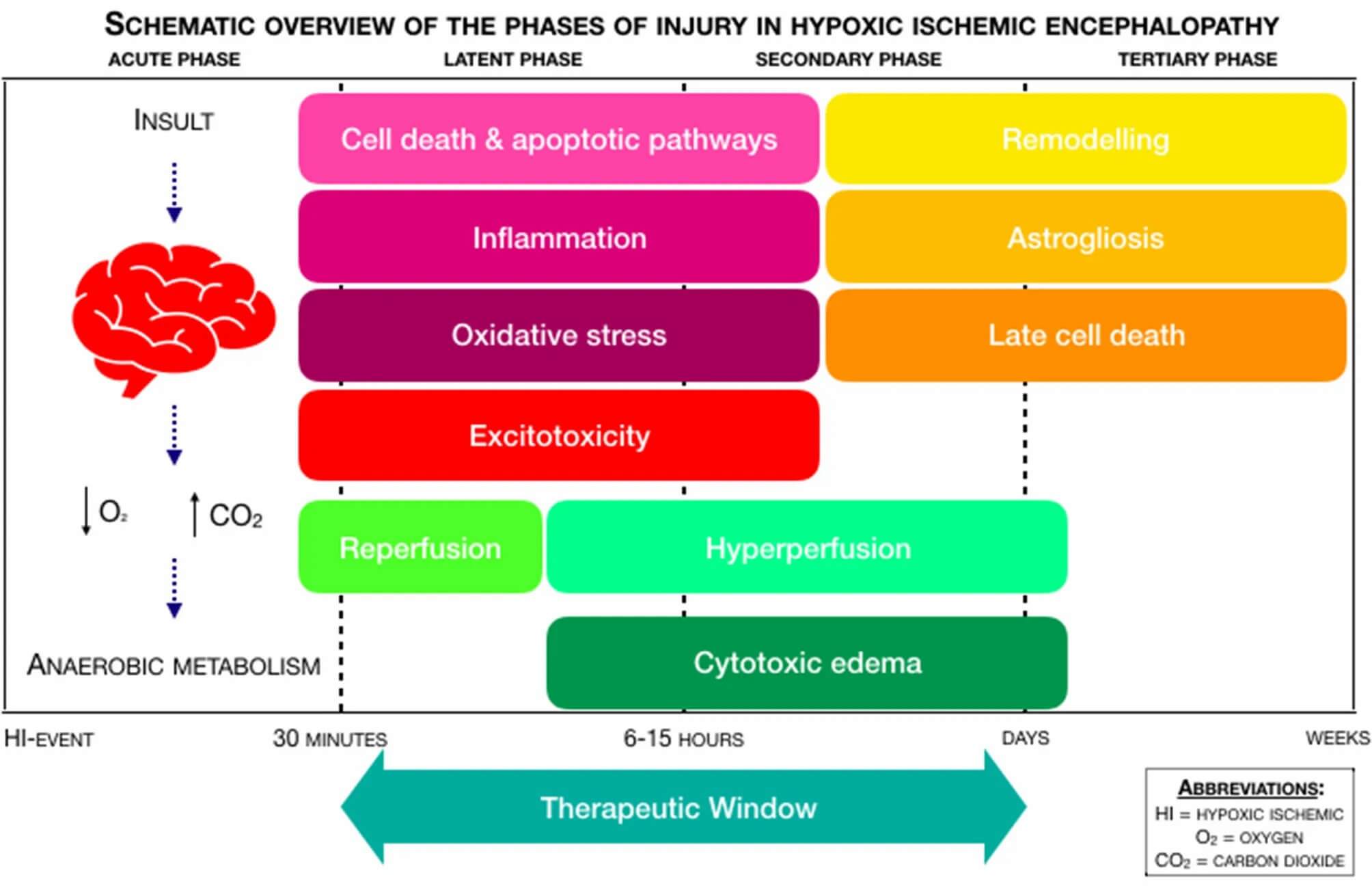 Fig. 1 Schematic overview of the phases of injury in Hypoxic-Ischemic Encephalopathy (HIE) (Kleushens D G, Costa F G, et al., 2021).
The current clinical management is largely dependent on therapeutic hypothermia which has a narrow therapeutic window of 6 hours and limited efficacy in severe cases. This unmet need highlights the importance of preclinical HIE models to identify synergistic therapies that may extend the treatment window or improve neuronal repair.
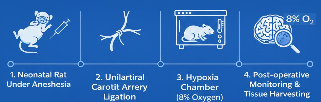
Validated HIE Animal Models
Our Rice–Vannucci HIE model is a widely accepted standard for neonatal hypoxic-ischemic brain injury research.
Model Principle:
Simulates the clinical pathology of neonatal asphyxia through unilateral permanent carotid artery occlusion combined with systemic hypoxia.
Animal Strains:
SD rats
Workflow
Comprehensive HIE Data Package
We provide a full-spectrum preclinical HIE model evaluation system for translational neuroscience research.
- Histological analysis
Nissl staining for neuronal integrity
TTC staining for cerebral infarct volume
H&E staining for histopathological evaluation of brain injury
TUNEL staining for detection of apoptotic neuronal cells
- Molecular biomarkers
Inflammatory cytokines: TNF-α, IL-1β, IL-6
Apoptosis markers: Caspase-3
Oxidative stress indicators
- Functional outcomes
Morris water maze (learning & memory)
Cylinder test (forelimb asymmetry)
Rotarod test (motor coordination and fatigue)
Study Examples
Cerebrovascular Dysfunction in Neonatal HIE Rats
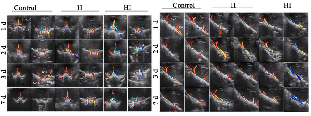 Fig. 2. TCD imaging revealed increased and reversed cerebral blood flow in ACA, MCA, and ICA-L in HI rats compared with controls, indicating significant cerebrovascular dysfunction after hypoxic-ischemic injury (Liu J X, Fang C L, et al., 2023).
Morphological Validation of HI-Induced Brain Injury
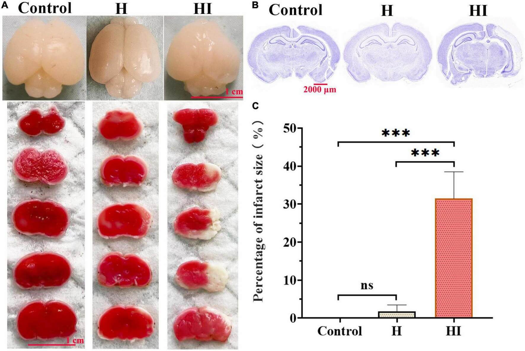 Fig. 3. TTC and Nissl staining demonstrated severe cerebral infarction, tissue atrophy, and neuronal loss in HI rats compared with control and hypoxia groups (Liu J X, Fang C L, et al., 2023).
Increased TNF-α Expression Following Hypoxic-Ischemic Injury
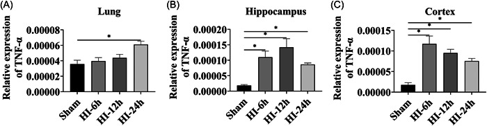 Fig. 4. RT-qPCR analysis showed significant upregulation of TNF-α in the lung, hippocampus, and cortex after HI injury, indicating a robust inflammatory response following hypoxic-ischemic insult (Niu YM, Du SZ, et al., 2023).
Cognitive Function Impairment Induced by HIE
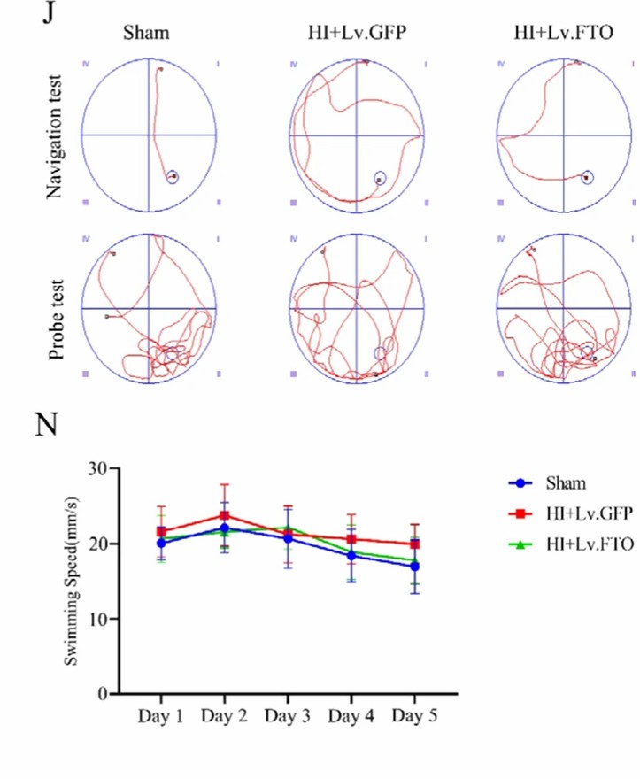 Fig. 5. Morris water maze analysis showed that HI rats exhibited impaired spatial learning and memory, while FTO overexpression significantly reduced escape latency and improved target quadrant retention and platform crossings (Deng, J., Liao, Y., et al., 2023).
Why Choose Creative Bioarray for HIE Research?

Exceptional Stability
Our optimized hypoxia conditions and surgical precision ensure experimental consistency and high model reliability.

Multi-Level Assessment
We don't just look at one metric. We integrate structural, molecular, and behavioral endpoints for a holistic drug assessment.

Surgical Expertise
Neonatal microsurgery is delicate. Our team's experience translates to higher survival rates and better data quality than industry averages.

Streamlined CRO Workflow
From initial study design to final data delivery, we maintain clear timelines and transparent reporting.
FAQ
Why is the P7 rat considered the "Gold Standard" for HIE?
The brain development of a P7 rat is roughly equivalent to a full-term human newborn. This makes it the most accurate window for studying neonatal asphyxia. We also offer P10 models for researchers targeting slightly more mature brain structures.
How do you handle the high mortality rate often associated with HIE surgery?
Precision is everything in neonatal surgery. We maintain strict thermal stability and customize hypoxia duration based on the specific animal strain to ensure survival rates that significantly exceed standard benchmarks.
What is the typical turnaround time for an HIE efficacy study?
A standard acute study (TTC/Biochemicals) typically takes 4–6 weeks, while long-term behavioral studies require 8–12 weeks.
Contact our team today to discuss your study design and see how our validated models can accelerate your program.
Request a project quote
References
- Niu YM, Du SZ, et al. TNF-α interference ameliorates brain damage in neonatal hypoxic-ischemic encephalopathy rats by regulating the expression of NT-3 and TRKC. Ibrain. 2023. 9(4):381-389
- Liu J-X, Fang C-L, et al. Transcranial Doppler Ultrasonography detection on cerebrovascular flow for evaluating neonatal hypoxic-ischemic encephalopathy modeling. Front. Neurosci. 2023. 17:962001.
- Deng, J., Liao, Y., et al. N6-methyladenosine demethylase FTO regulates synaptic and cognitive impairment by destabilizing PTEN mRNA in hypoxic-ischemic neonatal rats. Cell Death Dis. 2023.14, 820