What are the Differences Between M1 and M2 Macrophages?
What are Macrophages?
Macrophages are a type of white blood cells that play an important role in the human immune system. They carry out a variety of functions, including phagocytosis and digestion of microorganisms, removal of debris and dead cells, and stimulation of other cells involved in immune function. In addition to their immune role, macrophages also secrete anti-inflammatory cytokines and help mediate reparative processes.
Macrophages are derived from monocytes, which circulate through the blood for one to three days before migrating into tissues, where they become macrophages or dendritic cells. Macrophages can be found within many organs in the body, including the liver, brain, bones, and lungs, as well as in the blood, particularly at sites of infection.
What are the Types of Macrophages?
Activated macrophages with different phenotypes are usually classified into M1-macrophages (CAM) and M2- macrophages (AAM). The classically activated M1-macrophages comprise immune effector cells with an acute inflammatory phenotype. They are highly aggressive to bacteria and produce large amounts of lymphokines. The alternatively activated, anti-inflammatory M2-macrophages can be separated into at least three subgroups. These subtypes have a variety of different functions, including regulation of immunity, maintenance of tolerance and tissue repair/wound healing. Indeed, cells of the monocyte/macrophage lineage exhibit extraordinary plasticity in response to endogenous and exogenous stimuli, which can allow overriding of the initial M1/M2-polarization processes, for example M2-polarized macrophages can convert to the M1-activated status under certain conditions.
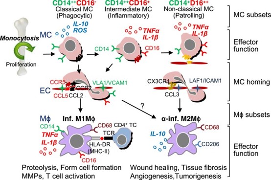 Human MC and M ϕ differentiation, and distinct subset functions (Yang, J., Zhang, L., Yu, C. et al., 2014).
The specific function of M1 macrophages is to detect, engulf and destroy bacteria. They can do this through phagocytosis, a process by which a bridge is formed between cellular receptors on the macrophage and surface antigens on the bacteria. Once the bridge is formed, the membrane of the macrophage protrudes out and surrounds the bacteria. Once inside the macrophage, the bacteria are trapped within a phagosome, or a vesicle, which then fuses with a lysosome. The lysosome contains enzymes and peroxides that are able to digest the pathogen.
M1 macrophages promote inflammation, extracellular matrix destruction, apoptosis of invading cells by releasing various cytokines and nitric oxide to aid in cellular destruction, and antigen presentation, making them antigen-presenting cells (APCs). After a macrophage processes and digests the antigen, a major histocompatibility complex (MHC) molecule delivers the antigen to the surface of the macrophage to allow a T-cell receptor to bind. This triggers the adaptive immune response, which recognizes the foreign antigen and mount further mechanisms to kill the cells.
M2 macrophages on the other hand are required for regeneration of connective tissue during wound healing. They produce vascular endothelial growth factor (VEGF) and transforming growth factor (TGF)-β1, which allows for vascular stability and wound repair. M2 macrophages also function to phagocytize bacteria and damaged tissue around the wound. They are then able to debride damaged tissue by releasing digestive enzymes such as proteases. Subsequently, they secrete growth factors that stimulate cells to re-epithelialize the wound, generate granulation tissue, and form new extracellular matrix.
Macrophages play additional roles in specific organ systems. For example, when air is inhaled into the lungs for oxygen exchange, toxic substances such as bacteria, viruses, and fungi may be taken into the lungs, especially the alveoli. Lung macrophages are able to process bacteria and other toxic substances in the alveoli to prevent diseases such as tuberculosis by forming granulomas. Within the liver, macrophages are called Kupffer cells. Kupffer cells clear the liver for potential pathogens that may enter the bloodstream from the gastrointestinal tract. They also play a role in maintaining iron levels and bilirubin metabolism. If the liver is injured, Kupffer cells secrete anti-inflammatory cytokines such as interleukin [IL]-10, IL-4, and IL-13. Macrophages of the brain are called microglia. Microglia have a neuroprotective effect and secrete a variety of anti-inflammatory cytokines and nerve injury factors. If neurons are damaged, microglia are recruited to the site, and can phagocytose dead cells and foreign materials to prevent further tissue damage.
Creative Bioarray Relevant Recommendations
Accelerate your programs with Creative Bioarray's extensive inventory and prospective network of macrophages.
| Cat. No. | Product Name |
| CSC-C4715Z | M1/M2 Macrophages |
| CSC-C4427X | Normal PB Monocyte Derived Macrophages |