Modern Histological Techniques
Modern histological methods have significantly improved our ability to study the biology of tissues and cells. They draw not just on traditional microscopic observations but also on molecular biology and computers to offer higher biological perspectives.
Microscopy Techniques
Optical microscopes
Optical microscopes serve as the main instruments used in histological analysis. Different types of microscopes such as compound light microscopes, fluorescence microscopes, and phase-contrast microscopes enable scientists to examine the large-scale structures of cells and tissues.
- Compound Light Microscope: It known as the compound light microscope employs multiple lenses to achieve magnification levels up to 1000x while maintaining a resolution of approximately 0.2 micrometers. The compound light microscope functions as an essential instrument to study cell morphology and tissue structures but lacks the resolution needed to visualize subcellular structures.
- Fluorescence Microscope: This microscope operates by activating fluorescent dyes in samples which emit light at specific wavelengths. The microscope generates high-contrast images which makes it ideal for studying specific molecular or ionic distributions within cells.
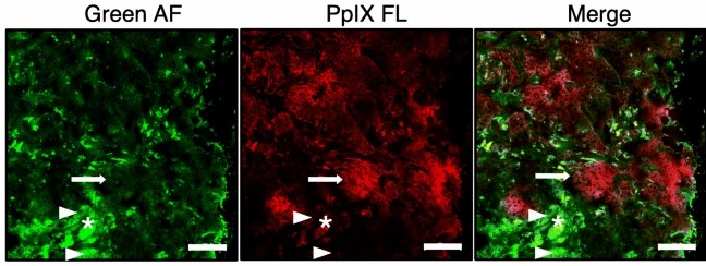 Fig. 1. Representative fluorescence microscopy of cancer cell-specific localization of ALA-induced PpIX (Ottolino-Perry K, Shahid A, et al., 2021).
- Confocal Microscope: The confocal microscope combines optical and fluorescence microscopy capabilities through precise laser beam focusing and scanning that eliminates stray light interference to generate high-resolution three-dimensional images with exceptional sensitivity. Scientists can simultaneously observe live cells and track molecular markers throughout different cellular layers with this method.
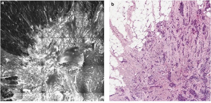 Fig. 2. Fluorescence confocal microscopy for invasive breast carcinoma (Ragazzi M, Piana S, et al., 2013).
Electron microscopes
By using electron beams instead of light beams electron microscopes achieve greater detail compared to conventional optical microscopes.
- Scanning electron microscopes (SEM): A scanning electron microscope produces images by directing an electron beam across a sample surface and detecting emitted secondary electrons. This microscope can magnify objects up to tens of thousands of times and reach a resolution of 1 nm which makes it perfect for examining surface morphology. SEM technology delivers exceptional performance for examining surface structures of cells in great detail.
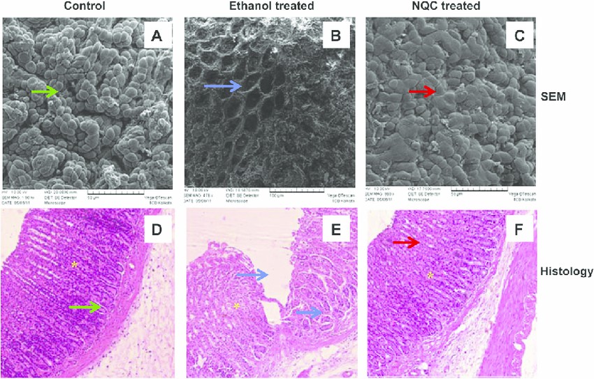 Fig. 3. Scanning electron microscopy (SEM) and histological analysis of rat gastric mucosa (Chakraborty S, Stalin S, et al., 2011).
- Transmission electron microscopes (TEM): Transmission electron microscopes create images by directing an electron beam through ultra-thin sample sections and analyzing the resulting electron scattering patterns. The instrument achieves a resolution of 0.1-0.2 nanometers and provides magnification up to hundreds of thousands of times which makes it ideal for examining cellular structures like mitochondria and ribosomes in detail. To operate the transmission electron microscope properly researchers must prepare precise ultrathin sections of samples which they then stain with heavy metals.
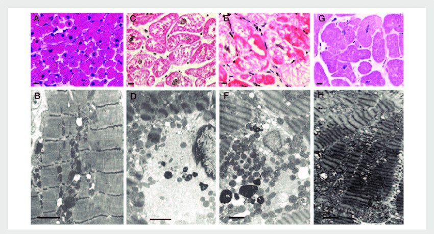 Fig. 4. Histology and transmission electron microscopy (TEM) of cardiomyopathy (Frustaci A, Sabbioni E, et al., 2012).
- Cryo-Electron Microscopy (Cryo-EM): Cryo-EM combines electron microscopy technology with cryogenic methods to produce three-dimensional images of biomolecules preserved in their quasi-natural conditions. Researchers must rapidly freeze samples to preserve their natural structure before capturing images using a low-temperature electron beam. The atomic-level resolution achieved by Cryo-EM makes it indispensable for revealing detailed structural information about proteins and viruses along with other biomolecules. It has wide applications in biomedical research.
Molecular Biology Techniques
- In situ hybridization: In situ hybridisation uses probes containing nucleic acids that can stick to the target DNA or RNA molecules and identify genes or transcripts within cells or tissues. Methods include fluorescent in situ hybridization (FISH) and radioactive in situ hybridization, both commonly used for gene mapping, chromosome anomaly detection and disease diagnosis.
- Immunohistochemistry and immunofluorescence: Immunohistochemistry (IHC) and immunofluorescence (IF) both use antibodies to label target proteins in tissues, allowing for the visualization of protein distribution and expression.
Image Analysis Techniques
Photomicroscopy, combined with image analysis, provides more quantitative and qualitative data for histology.
Computer-assisted image analysis
Computer-aided image analysis technology utilizes computer vision, deep learning, and machine learning algorithms to process and analyze medical images, thereby enhancing diagnostic accuracy and efficiency. Its main applications include:
- Image Segmentation and Feature Extraction: Deep learning models like convolutional neural networks (CNNs) enable this technology to segment and identify cells, tissue structures and lesion areas in images which supports quantitative analysis.
- Image Enhancement and Fusion: By adjusting parameters such as contrast and brightness, image quality is optimized, allowing researchers to observe fine structures more clearly.
- Automated Diagnostic Support: Deep learning algorithms enable computerized image analysis systems to quickly process massive datasets while minimizing human errors and enhancing diagnostic performance.
Three-dimensional reconstruction
Software aggregates and analyses multiple images from two dimensions to recreate the three-dimensional geometry of a specimen, offering a more intuitive visualization of the spatial shape of cells and tissues. This approach is particularly useful in developmental biology and neuroscience because it enables 3D reconstruction of entire organ or cell structure.
- Three-Dimensional Reconstruction of Histological Sections: This technology uses consecutive section scanning techniques along with computer algorithms to transform two-dimensional section data into three-dimensional models that faithfully replicate internal tissue structures while supporting biological and medical research.
- Three-Dimensional Reconstruction of Medical Imaging: Imaging methods such as CT and MRI scanning produce two-dimensional images that software algorithms transform into accurate 3D models through three-dimensional reconstruction techniques. These models permit examination of lesion areas from multiple angles which supports surgical planning and simulation activities.
Tissue Arrays
Tissue arrays provide high-throughput technology to look at many tissue samples at a time. By strategically stacking several small tissue sections (usually from different people, patients or experimental teams) on a microscope slide, researchers can then analyze them all in parallel under the same conditions — resulting in a marked increase in experimental productivity and data integrity.
Creative Bioarray Relevant Recommendations
| Products & Services | Description |
| Transmission Electron Microscopy (TEM) Service | Creative Bioarray has scientists and imaging laboratories to perform the full comprehensive Transmission Electron Microscopy (TEM) and Scanning Transmission Electron Microscopy (STEM) Service for the biological sciences and clinical research including plant samples, animal samples, bacteria, and pathology specimens. |
| Scanning Electron Microscopy (SEM) | Creative Bioarray is dedicated to providing high-quality SEM services tailored to meet your specific needs. |
| Cryogenic Transmission Electron Microscopy (cryoTEM) | Creative Bioarray provides Cryogenic Transmission Electron Microscopy (cryoTEM) for you. |
References
- Ragazzi, M., Piana, S., et al. Fluorescence confocal microscopy for pathologists. Mod Pathol. 2014. 27, 460–471.
- Ottolino-Perry, K., Shahid, A., et al. Intraoperative fluorescence imaging with aminolevulinic acid detects grossly occult breast cancer: a phase II randomized controlled trial. Breast Cancer Res. 2021. 23, 72.
- Chakraborty S, Stalin S, et al. The use of nano-quercetin to arrest mitochondrial damage and MMP-9 upregulation during prevention of gastric inflammation induced by ethanol in rat. Biomaterials. 2012 Apr;33(10):2991-3001.
- Frustaci A, Sabbioni E, et al. Selenium- and zinc-deficient cardiomyopathy in human intestinal malabsorption: preliminary results of selenium/zinc infusion. Eur J Heart Fail. 2012 Feb;14(2):202-10.