Pulmonary Fibrosis Models
Creative Bioarray offers well-characterized pulmonary fibrosis animal models to support preclinical drug efficacy evaluation, mechanistic research, and translational decision making. Our portfolio includes well characterized models of idiopathic pulmonary fibrosis (IPF) and other fibrotic interstitial lung diseases, enabling clients to assess therapeutic candidates across key biological endpoints — from inflammation and fibroblast activation to extracellular matrix (ECM) deposition and impaired lung function.
- Background
- Models
- Study Examples
- Features
- FAQ
Overview of Pulmonary Fibrosis
Pulmonary fibrosis is a subset of interstitial lung disease that is characterized by the scarring of lung tissue which leads to difficulty breathing. Idiopathic pulmonary fibrosis (IPF) is one of the most common types of pulmonary fibrosis and is seen most frequently in North America and Europe. The reported incidence of IPF is approximately 3–9 cases per 100,000 people each year.
Pulmonary fibrosis involves the activation of fibroblasts in the lungs which leads to excess deposition of ECM protein and consequent tissue remodeling and scarring. The fibrotic process is understood to occur in three stages. The initial stage is alveolar epithelial injury, which can be caused by environmental pollutants, drugs, radiation, genetic predisposition, or unknown factors. Injury to epithelial cells causes a localized inflammatory response which recruits various immune cells to the site of injury and results in secretion of cytokines. During fibroblast activation and proliferation, which is thought to be predominantly driven by TGF β/Smad signaling, fibroblasts transition into myofibroblasts which deposit large amounts of ECM proteins including collagen.
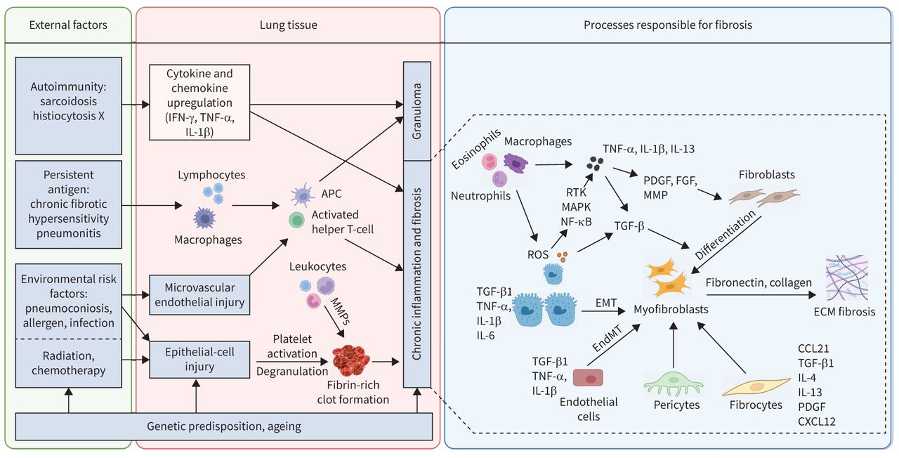 Fig. 1. Pathogenesis of Pulmonary Fibrosis: Cascade from Lung Injury to Fibrosis (Kolanko E, et al., 2023).
Fibrosis is irreversible which makes the disease difficult to treat. While there are currently FDA approved antifibrotic therapies like pirfenidone and nintedanib, these medications can only slow disease progression—they cannot reverse the symptoms of pulmonary fibrosis or improve lung function. This highlights the urgent need for preclinical PF models that faithfully mimic human disease, to enable the discovery of truly effective treatments.
Creative Bioarray's Pulmonary Fibrosis Models
Bleomycin-Induced Pulmonary Fibrosis (Standard and Enhanced Models)
Animal Strain: C57BL/6 mice (6-8 weeks, male or female)
Induction Method: Single or repeated intratracheal instillation, or oropharyngeal/nebulized administration of bleomycin sulfate, inducing progressive pulmonary fibrosis.
Disease Relevance: This combined Bleomycin approach allows flexible dosing and administration routes, improving reproducibility, uniform lung injury, and persistence of fibrosis for both acute and chronic studies. It remains the gold standard preclinical model for evaluating anti-fibrotic drug candidates.
Silica-Induced Pulmonary Fibrosis
Animal Strain: C57BL/6 mice
Induction Method: Intratracheal instillation of crystalline silica particles to simulate chronic environmental exposure related fibrosis.
Disease Relevance: This model mimics non-resolving fibrosis, such as silicosis, providing complementary insights for compounds targeting chronic fibrotic processes.
Endpoints:
Histopathological Evaluation
- Masson's trichrome staining
- Collagen deposition analysis
- Alveolar structure injury assessment
- Inflammatory cell infiltration evaluation
- Granuloma formation analysis
Biochemical and Molecular Analysis
- Lung hydroxyproline quantification
- BALF inflammatory cytokine detection
- Fibrosis-related cytokine analysis, including TGF-β1 and IL-6
- TNF-α and additional profibrotic biomarker assessment
Pulmonary Function Assessment (Optional)
- Pulmonary compliance measurement
- Respiratory mechanics evaluation
- Airway resistance assessment
- Lung function impairment analysis
Study Examples
Lung Fibrosis Imaging
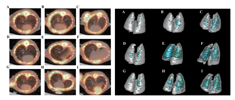 Fig. 3. PET/CT and 3D ROI images at Day 25–26 show increased non‑aerated lung volume in bleomycin-treated mice vs saline controls, indicating progressive fibrosis. A IPC, D ITC, G IVC are those in saline administration controls, BLM lower dose groups B IPL, E ITL, H IVL and high dose groups C IPH, F ITH, I IVH, showed an increase in non-aerated lung area (Gul A, et al., 2023).
Biomarker Changes
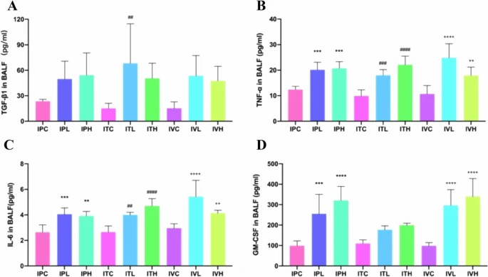 Fig. 4. Bar charts show elevated TGF‑β1, TNF‑α, IL‑6, GM‑CSF in BALF/serum and increased hydroxyproline/PAI‑1 in lung tissue of bleomycin-treated mice, confirming fibrosis and ECM deposition (Gul A, et al., 2023).
Lung Histopathology
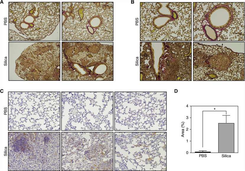 Fig. 5. Silica-administered mouse lungs showed severe inflammatory cell accumulation in alveoli with partial, focal fibrosis (A, B). α-SMA staining revealed significantly expanded myofibroblast activation areas (C, D). These results confirm that single silica challenge successfully induced lung inflammation and fibrosis persisting for 24 weeks (Sugimoto N, et al., 2019).
Why Choose Creative Bioarray

Deep Disease Modeling Expertise
Our team has years of experience creating and validating predictive disease models tailored to your drug's mechanism of action.

Robust Data Quality & Analysis
We combine histopathology, biochemistry, and functional readouts to provide comprehensive efficacy profiles that support IND enabling decisions.

Customizable Study Designs
Whether you are in the early stages of proof of concept or advanced stages of preclinical candidate screening, we have flexible protocols and multiple dosing strategies to fit your needs.

Translational Focus
Our models emphasize clinical relevancy and mechanistic insight, increasing confidence in therapeutic translation.
FAQ
Q: What makes Creative Bioarray's bleomycin model a good choice for pulmonary fibrosis?
A: Our bleomycin model reproducibly induces many of the pathological features present in human pulmonary fibrosis. Bleomycin instillation results in injury to epithelial cells, infiltration of inflammatory cells, accumulation of collagen, and disturbed architecture. Due to this, bleomycin is one of the most popular animal models of pulmonary fibrosis used in preclinical studies.
Q: Can these models be used to better model chronic fibrosis or acute fibrosis?
A: Yes! By varying the dose and route of administration, you can tailor the model to your needs. Acute fibrosis can be modeled by a single intratracheal instillation of bleomycin, while chronic fibrosis can be modeled by repeated instillation or nebulization.
Ready To Get Started?
Explore effective pulmonary fibrosis models and gain high quality data to propel your drug discovery process. Contact us today to speak to one of our scientists and receive a custom study brochure.
References
- Kolanko E, Carganoni A, et al. The evolution of in vitro models of lung fibrosis: promising prospects for drug discovery. European Respiratory Review. 2024. 33(171): 230127;
- Gul A, Yang F, et al. Pulmonary fibrosis model of mice induced by different administration methods of bleomycin. BMC Pulm Med. 2023. 23, 91.
- Sugimoto N, Suzukawa M, et al. IL-9 Blockade Suppresses Silica-induced Lung Inflammation and Fibrosis in Mice. Am J Respir Cell Mol Biol. 2019. 60(2):232-243.