

Human Pancreatic Cancer Associated Fibroblasts
Cat.No.: CSC-C30031J
Species: Human
Source: Pancreas
Cell Type: Fibroblast
- Specification
- Background
- Scientific Data
- Q & A
- Customer Review
Pancreatic cancer-associated fibroblasts (CAFs) exhibit remarkable phenotypic and functional heterogeneity. Single-cell transcriptomic analyses have identified at least three major CAF subtypes in PDAC: myofibroblastic CAFs (myCAFs), inflammatory CAFs (iCAFs), and antigen-presenting CAFs (apCAFs). More recent integrated analyses have further refined this landscape, revealing nine distinct CAF subtypes including transitional CAFs (tCAFs), proliferative CAFs (pCAFs), and CDCP1⁺FTL⁺ CAFs. These subtypes exhibit differentiation plasticity, transitioning from early normal-like CAFs to iCAFs and myCAFs, ultimately giving rise to more invasive pCAFs.
The functional significance of pancreatic CAFs extends across multiple pro-tumorigenic axes: remodeling the extracellular matrix, secreting growth factors and cytokines such as TGF-β and IL-6, promoting angiogenesis, and establishing an immunosuppressive niche. Notably, CAFs can either promote or suppress tumor progression in a context-dependent manner. Specific subtypes, including ECM-remodeling myCAFs, hypoxic CAFs, and iCAF_chemokine subpopulations, have been associated with immune-excluded, chemoresistant phenotypes and poor clinical outcomes. Furthermore, CDCP1⁺FTL⁺ CAFs exhibit enhanced glycolysis and iron metabolism, resisting ferroptosis.
Human pancreatic CAFs are invaluable as a physiologically relevant in vitro model system. These cells are extensively employed in co-culture systems with PDAC cell lines and patient-derived organoids to recapitulate tumor-stroma interactions, study CAF-mediated chemoresistance, and evaluate novel therapeutic interventions. CAF-related gene signatures have demonstrated robust prognostic value, effectively stratifying patient survival and predicting treatment response. Collectively, human pancreatic CAFs are indispensable for dissecting PDAC pathogenesis and advancing precision oncology, serving as both a powerful experimental platform and a compelling therapeutic target.
Cancer-Associated Fibroblast-Derived MMP11 Promotes Tumor Progression in Pancreatic Cancer
Matrix metalloproteinase 11 (MMP11), a zinc-dependent endopeptidase involved in extracellular matrix degradation and remodeling, has been identified as a tumor promoter in multiple cancer types. However, its expression pattern and role in pancreatic ductal adenocarcinoma (PDAC) remain unclear. In this study, elevated MMP11 expression was identified in PDAC tissues and was associated with diminished survival. Integrated single-cell RNA sequencing and co-immunofluorescence staining revealed that MMP11 was predominantly expressed in cancer-associated fibroblasts (CAFs).
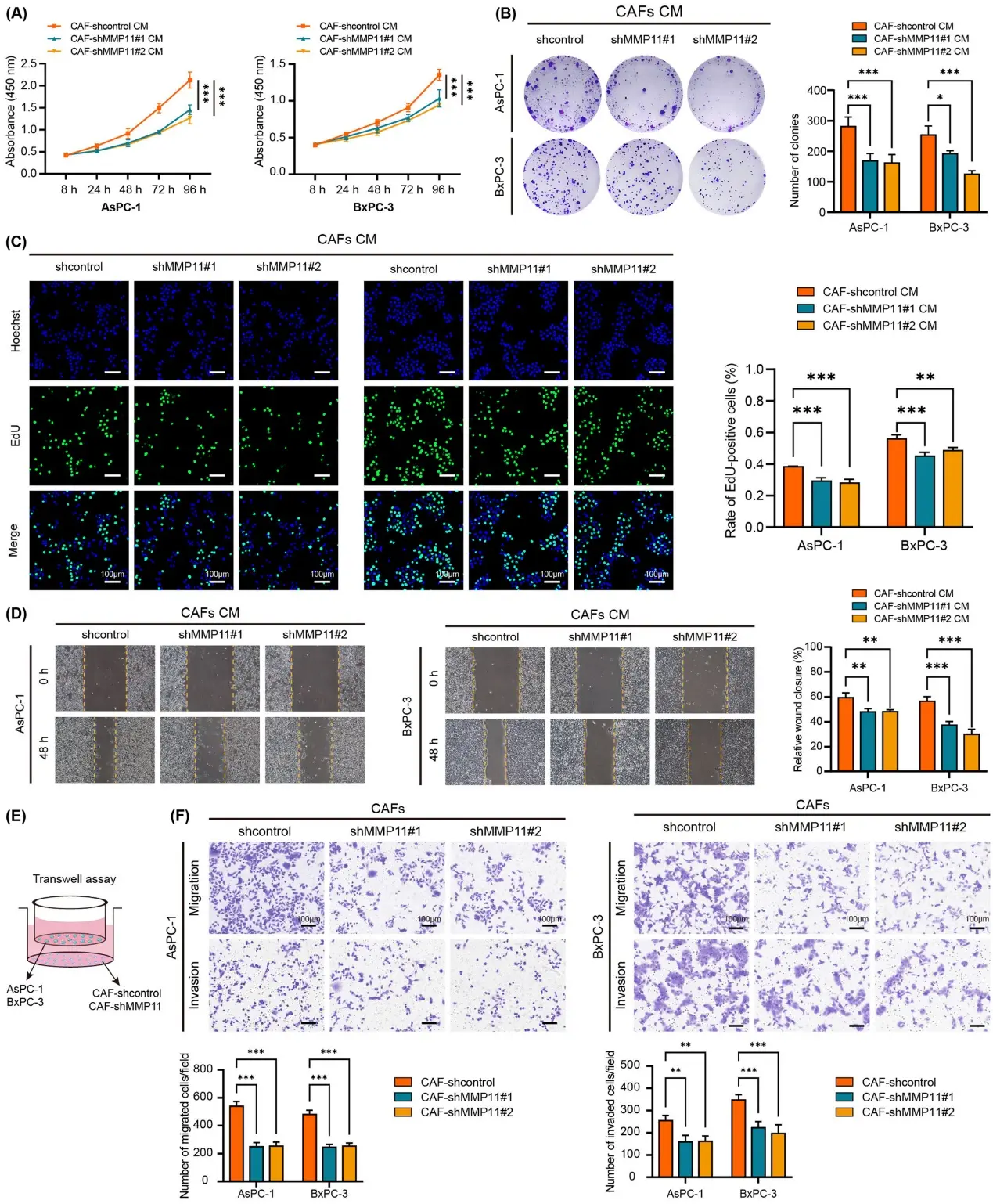
To elucidate the potential role of CAF-derived MMP11 in PDAC progression, we generated stable MMP11-knockdown CAFs using lentiviral vectors carrying shRNA against MMP11 or control. Subsequently, conditioned medium (CM) was collected from CAF-shcontrol and CAF-shMMP11 and incubated with AsPC-1 and BxPC-3 cells. CCK-8 assay demonstrated that the proliferative capacities of AsPC-1 and BxPC-3 cells exposed to CM from CAF-shMMP11 were markedly impaired compared with CM from CAF-shcontrol (Fig. 1A). Colony-formation assays (Fig. 1B) and EdU assays (Fig. 1C) showed similar results. Furthermore, we explored the effect of CAF-derived MMP11 on PDAC cell migration and invasion. Wound-healing assays showed that CM from CAF-shMMP11 significantly inhibited the migration of AsPC-1 or BxPC-3 cells compared with CM from CAF-shcontrol (Fig. 1D). In transwell assays, stable MMP11-knockdown or control CAFs were seeded into the lower chamber and PDAC cells (AsPC-1 and BxPC-3) were seeded into the upper chamber (Fig. 1E). We found that the number of migrated and invaded cells was reduced by co-culture with CAF-shMMP11 compared with the CAF-shcontrol (Fig. 1F).
Ask a Question
Write your own review
Description: Human Pancreatic Microvascular Endothelial Cells from Creative Bioarray are isolated from human pancreatic tissue. Human Pancreatic Microvascular Endothelial Cells are grown in T25 tissue culture ...
Description: Pancreatic Stellate Cells (HPaSteC) are the main fibroblastic cells of the pancreas. HPaSteC are responsible for the synthesis and the degradation of the extracellular matrix proteins that promote ...
Description: Primary Human Pancreatic Stromal Cells were initiated by elutriation from normal human pancreatic tissue.These cells were originated using Complete Serum-Free Medium Kit With SuperFuel™, are ...
Description: Human Pancreatic Islets are isolated from the pancreas of human using Collagenase P and purified using Ficoll density gradient.
Description: Human Primary Pancreatic Fibroblasts are isolated from normal human pancreatic tissue. Human Primary Pancreatic Fibroblasts are grown in T75 tissue culture flasks pre-coated with gelatin-based ...
Description: Human Pancreatic Epithelial Cells from Creative Bioarray are isolated from normal human pancreatic tissue. Human Pancreatic Epithelial Cells are grown in T25 tissue culture flasks pre-coated with ...