Comparison of Different Methods to Measure Cell Viability
In vitro assessment of cellular viability has become a generic approach in addressing a vast range of biological questions in many areas of biomedical research. Cell viability assays are often used to screen collections of compounds to determine if the test molecules have effects on cell proliferation or show direct cytotoxic effects that eventually lead to cell death. According to the number and type of cells used, choosing a proper assay method to decided whether the expected outcome is valid or not.
Viable cells can convert the substrate into product resulting in a colored or fluorescent product that can be detected with a plate reader, and the signal is directly proportional to the number of viable cells present. But the dying cells lose the conversion ability, that difference provides the basis for many of the commonly used cell viability assays. The tetrazolium reduction, resazurin reduction, and protease activity assays all take advantage of this theory. The ATP assay is somewhat different in that the addition of assay reagent immediately ruptures the cells, thus there is no incubation period of the reagent with a viable cell population.
MTT Tetrazolium Assay
Principle
The MTT tetrazolium reduction assay was the first homogeneous cell viability assay developed for a 96-well format that was suitable for high throughput screening (HTS).
NAD(P)H-dependent cellular oxidoreductase enzymes in viable cells can reduce the tetrazolium dye MTT 3-(4,5-dimethylthiazol-2-yl)-2,5-diphenyltetrazolium bromide to its insoluble formazan, which has a purple color. The quantity of formazan is measured by recording changes in absorbance at 570nm using a spectrophotometer. Under defined conditions, it can reflect the number of viable cells present. MTT assays can be used to measure cytotoxicity (loss of viable cells) or cytostatic activity (shift from proliferation to quiescence) of potential medicinal agents and toxic materials.
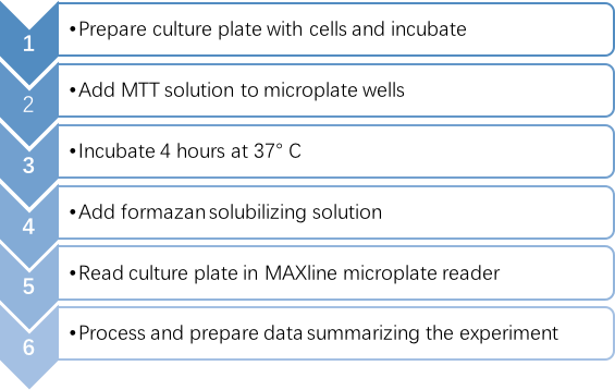 Figure 1. The process of the MTT colorimetric assay.
 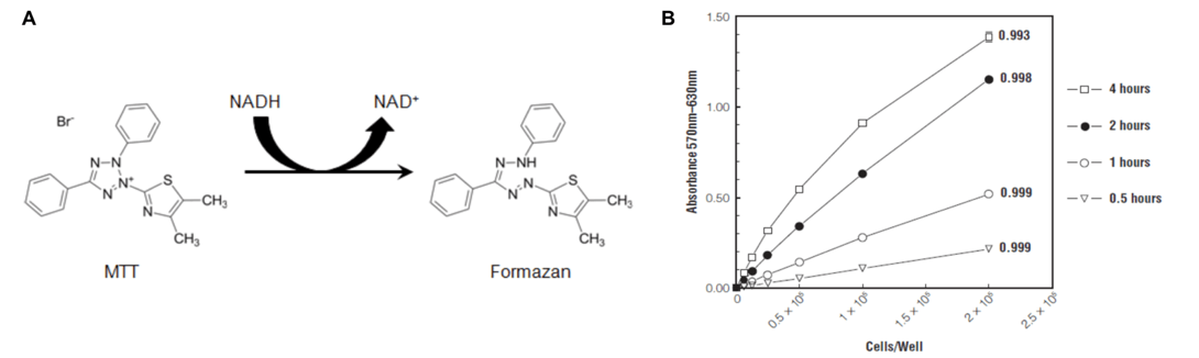 Figure 2. A: Structures of MTT and colored formazan product. B: Direct correlation of formazan absorbance with B9 hybridoma cell number and time-dependent increase in absorbance (Riss, T. L. 2004).
Advantages
- Fewer steps, uses fewer materials, and does not carry the added burden of radioactive waste disposal.
Disadvantages
- Not suitable for suspending cells.
- Must be optimized for seeding amount and assay duration to obtain satisfactory results.
- Precipitated protein and cellular debris present in the culture plate wells are a potential source of experimental error because they interfere with the optical readings.
MTS Tetrazolium Assay
Principle
MTS [3-(4,5-dimethylthiazol-2-yl)-5-(3-carboxymethoxyphenyl)-2-(4-sulfophenyl)-2H-tetrazolium] is a new kind of tetrazolium salts, it can be reduced by viable cells to generate formazan products that are directly soluble in cell culture medium. For these improved tetrazolium reagents, the usual protocol is simply to add them to the culture medium and read the absorption of the reduced product at an appropriate time. This approach eliminates a liquid handling step during the assay procedure, thus it obviously saves time and eliminates potential errors such as cell loss that can occur when removing culture medium and subsequently solubilizing cells.
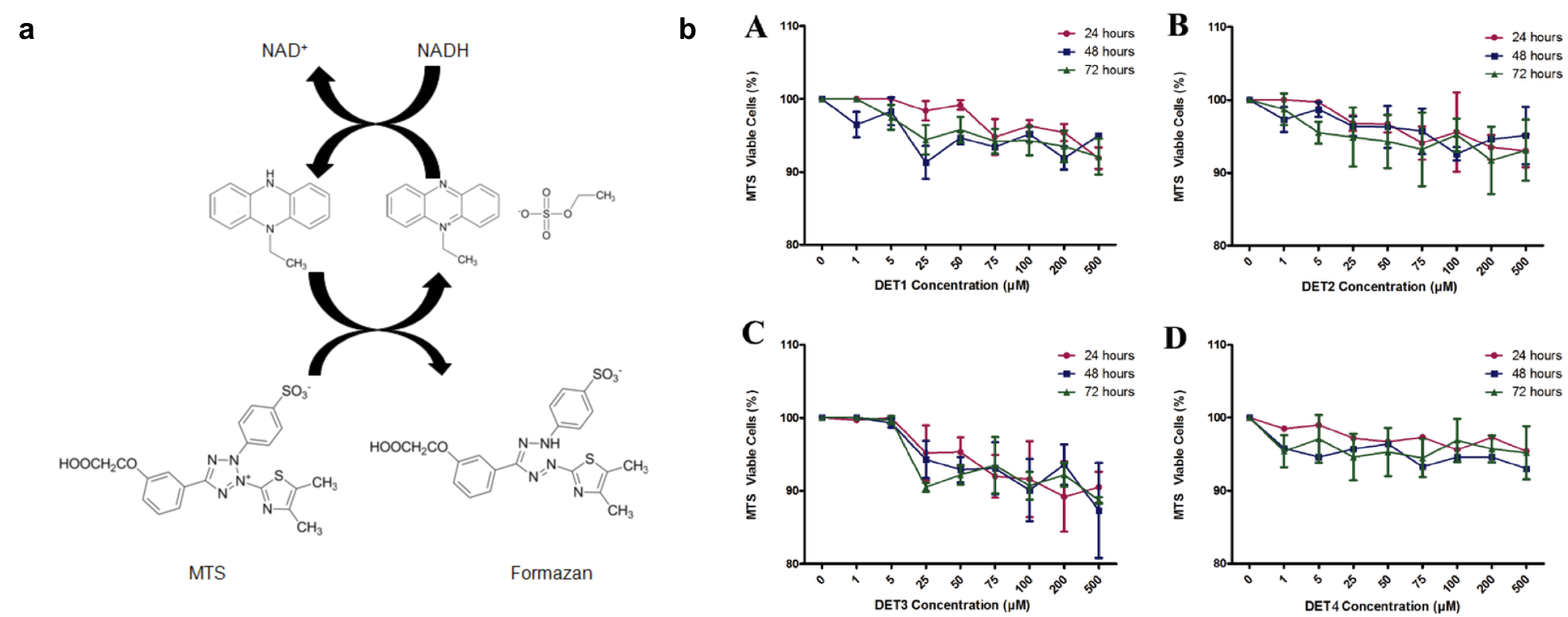 Figure 3. a: Intermediate electron acceptor pheazine ethyl sylfate (PES) transfers electron from NADH in the cytoplasm to reduce MTS in the culture medium into an aqueous soluble formazan. b: MTS assay for cell viability was performed with increasing concentration of different peptides at different time courses. (Alhoot, 2013)
Advantages
- Simple operation. MTS is more efficient than MTT and produces water-soluble formazan the does not require DMSO dissolution.
- Due to the MTS produce darker formazan product, the absorbance value range is more sensitive and accurate, as well as it's easier to judge positive results.
- Fast. 2-3 hours are enough for reaction while the MTT need 4 hours.
- Good repeatability.
Disadvantages
- The MTS regents is more expensive than MTT.
CCK-8 (WST-8) Assay
Principle
As a second-generation water-soluble tetrazolium salt, WST-8 [2-(2-methoxy-4-nitrophenyl)-3-(4-nitrophenyl)- 5-(2,4-disulfophenyl)-2H-tetrazolium, monosodium salt] has better soluble than MTS. WST-8 is reduced by dehydrogenases in cells to give an orange colored product (formazan), which is soluble in the tissue culture medium. And the amount of the formazan dye generated by dehydrogenases in cells is directly proportional to the number of living cells.
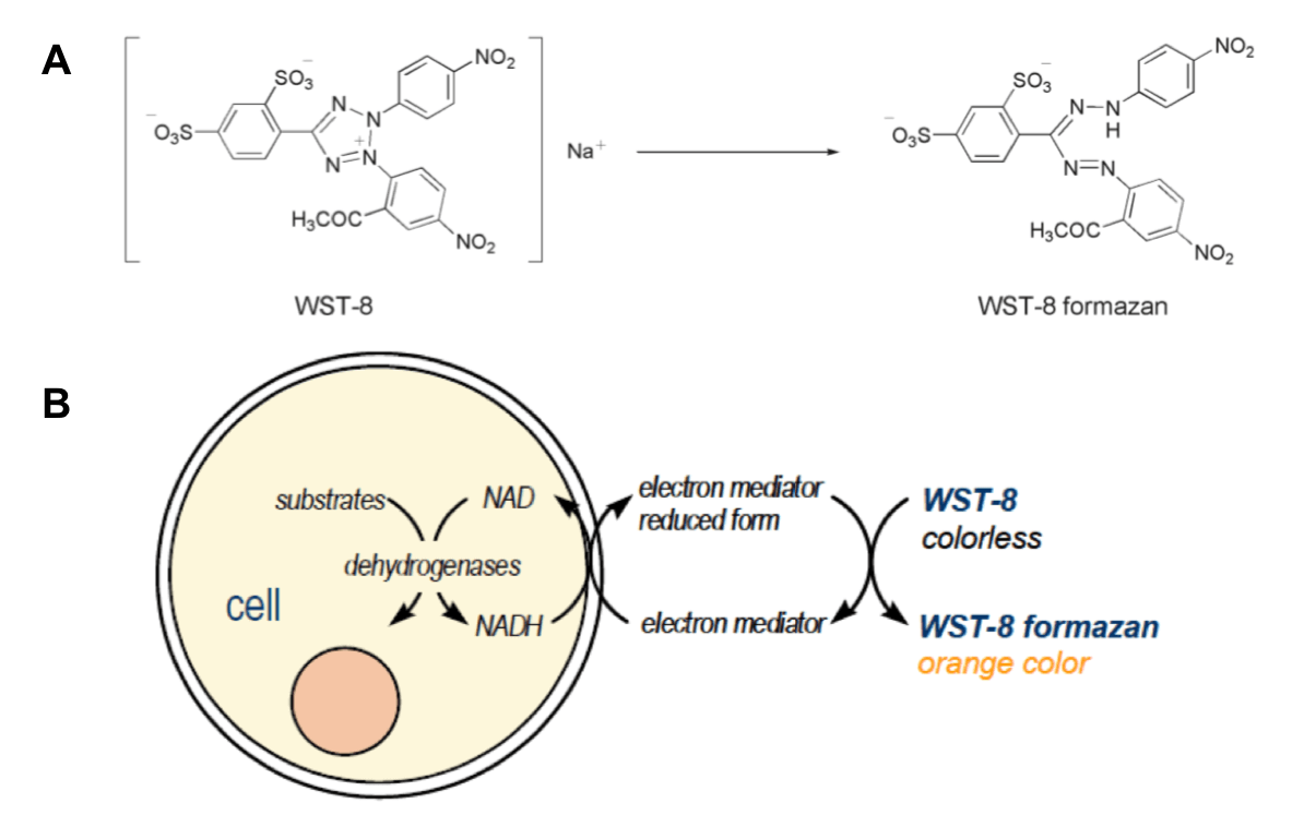
Figure 4. A: Structures of WST-8 and WST-8 formazan. B: Principle of the cell viability detection with Cell Counting Kit-8.
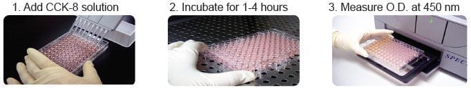
Figure 5. The procedure of CCK-8 assay.
Advantages
- CCK-8 is a one-bottle solution; no premixing of components is required.
- CCK-8 is simple, rapid, low consumed and sensitive with repeatability.
- CCK-8, being nonradioactive, allows sensitive colorimetric assays for the determination of the number of viable cells in cell proliferation and cytotoxicity assays.
Disadvantages
- More expensive than MTT regents
- the color of CCK-8 is closed to the medium with phenol red which may miss adding regents if not careful. The operation mistake will disturb the data result.
Sulforhodamine B colorimetric assay
Principle
The sulforhodamine B (SRB) assay was developed in 1990, one of the most widely used methods for in vitro cytotoxicity screening. The assay relies on the ability of SRB to bind to protein components of cells: SRB is a bright-pink aminoxanthene dye with two sulfonic groups that bind to basic amino-acid residues components of cells under mildly acidic conditions, and dissociate under basic conditions. As the binding of SRB is stoichiometric, the amount of dye extracted from stained cells can be used as a proxy for cell mass directly, and the amount of bound dye is proportional to the cell mass.

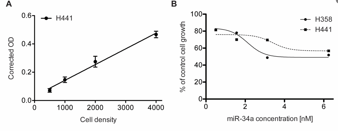
Figure 6. Sulforhodamine B colorimetric assay in cell culture to analyze cell proliferation. A. Cell number titration (H441) using the SRB assay 384-well format, n = 6, error bars = SD. B. miR-34a dose response analysis in lung cancer cell lines H441 and H358 using SRB 96-well format, n = 3. (Vichai, V, 2006)
Advantages
- Because the cell was fixed, SRB staining rarely affected by other compounds interfere which can remain for a long time.
- SRB soluble with Tris-base solution is also stable for a long time. Therefore, the 96-hole cell culture plate at different time points can be determined at the same time.
Disadvantages
- The application of the SRB assay is limited to manual or semiautomatic screening due to the multiple washing and drying steps, which are not amenable to automation.
- The operation procedure is complicated.
Conclusion
| MTT | MTS | CCK-8 | SRB | |
| Solubility | Indissolvable | Water soluble | Water soluble | Indissolvable |
| Detection Wavelength | 490nm | 450nm | 450nm | 510nm |
| Character | Solid | Liquid | Liquid | Liquid |
| Usage | Prepare the solutions | Use it right after it was ready | No need to prepare | Prepared beforehand |
| Need to redissolve or not | Yes, by DMSO | No | No | Yes, by Tris-base solution |
| Convenience | ++ | +++ | +++ | + |
| Detection speed | + | ++ | +++ | + |
| Repeatability | + | ++ | +++ | ++ |
| Stability | + | + | ++ | +++ |
Related Products & Services
Cell Viability Assays
Cell Proliferation Kit (MTT)
MTS Cell Proliferation Colorimetric Assay Kit
BrdU Cell Proliferation Assay Kit
BrdU Staining Kit for Flow Cytometry FITC
References
- Riss, T. L.; et al. Assay guidance manual. Sittampalam, GS, et al (2004).
- Alhoot, M. A.; et al. Inhibition of dengue virus entry into target cells using synthetic antiviral peptides. International journal of medical sciences. 2013, 10(6), 719.
- Vichai, V.; et al. (2006). Sulforhodamine B colorimetric assay for cytotoxicity screening. Nat Protoc. 2006, 1(3): 1112-1116.