Experimental Autoimmune Uveitis (EAU) Model
- Background
- Models
- Study Examples
- Features
- FAQ
Creative Bioarray provides validated Experimental Autoimmune Uveitis (EAU) models for preclinical ophthalmology research and autoimmune disease drug development. Our in vivo EAU platforms support efficacy evaluation, mechanism studies, and translational assessment of anti-inflammatory and immunomodulatory therapies with comprehensive functional, pathological, and immune-related analyses.
Overview of Autoimmune Uveitis
Non-infectious autoimmune uveitis is one of the biggest causes of blindness globally and remains difficult to manage clinically due to the recurrent inflammation and long-term treatment toxicity. Current therapy with corticosteroids and systemic immunosuppressants can decrease the inflammation but are commonly linked with recurrence, cataract, glaucoma, and systemic side effects. Although biologics such as anti-TNF medicines have improved outcomes in a subset of patients, therapy resistance and inadequate disease management remain common.
Autoimmune uveitis is largely mediated by abnormal T cell responses to retinal antigens. The activated Th1 and Th17 cells secrete inflammatory cytokines such as IFN-γ, IL-17 and TNF-α, which cause the disintegration of the blood-retina barrier, the influx of immune cells, retinal degeneration and visual impairment.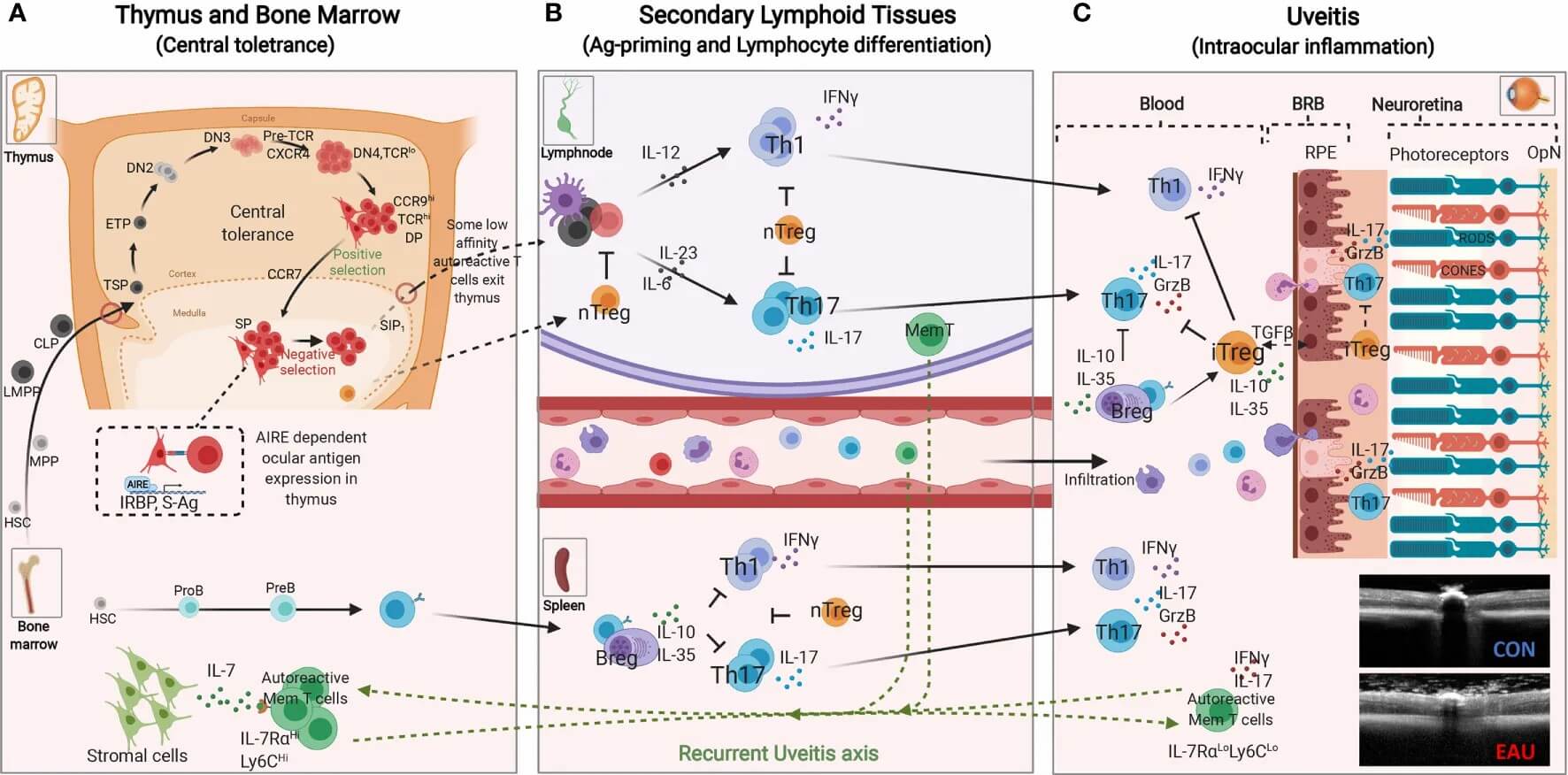
Fig. 1. Immunopathogenic Mechanisms of experimental autoimmune uveitis (EAU) (Egwuagu C E, Alhakeem S A, et al., 2021).
The complexity of human ocular immune responses mandates the continued use of translational animal models to understand disease development and to evaluate potential treatments. Experimental Autoimmune Uveitis (EAU) models accurately mimic the pathogenic and immunological aspects of human posterior uveitis and are widely employed in preclinical drug discovery.
Our Experimental Autoimmune Uveitis (EAU) Animal Model
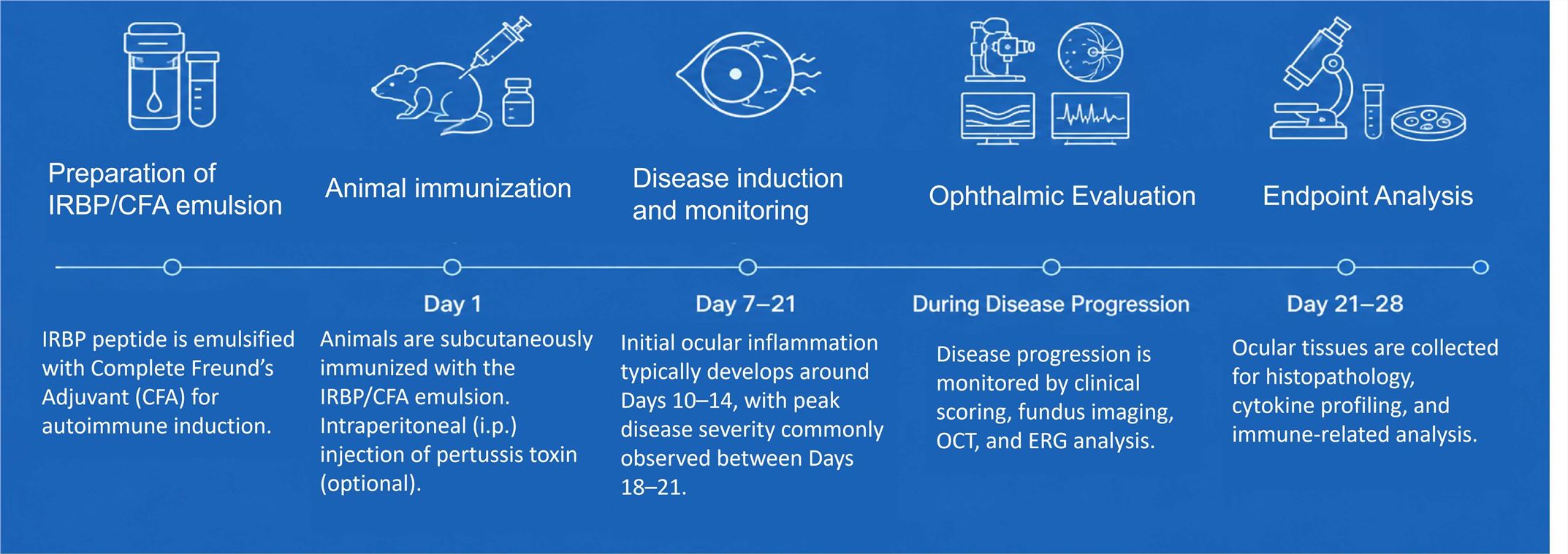
At Creative Bioarray, Experimental Autoimmune Uveitis (EAU) models are established by immunization with retinal autoantigens such as IRBP peptides emulsified in Complete Freund's Adjuvant (CFA), with optional pertussis toxin administration depending on the animal strain and study objective.
This induction strategy triggers autoreactive T cell responses dominated by Th1 and Th17 pathways, leading to blood-retina barrier disruption, inflammatory cell infiltration, retinal injury, and visual dysfunction that closely resemble human autoimmune posterior uveitis.
Animal Strains
- C57BL/6 mice
- B10.RIII mice
Evaluation Endpoints
Functional and Imaging Endpoints
- Clinical ophthalmic scoring
- Fundoscopy
- Optical coherence tomography (OCT)
Histopathology and Immunology
- Retinal inflammatory infiltration
- Histopathological scoring
- Cytokine analysis (IL-17, IFN-γ, TNF-α)
- Immune cell profiling
Study Examples
Retinal Inflammation and Structural Damage in IRBP-Induced EAU Mice
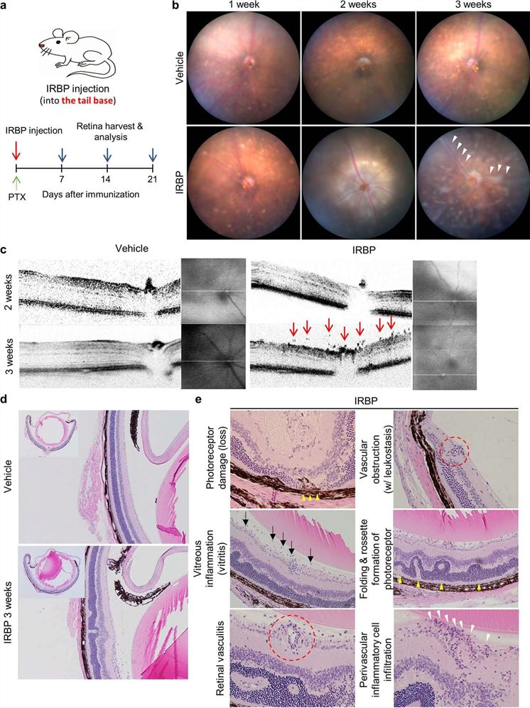 Fig. 2. IRBP-immunized mice developed characteristic features of autoimmune uveitis, including retinal vasculitis, inflammatory cell infiltration, vascular sheathing, and retinal structural disruption, as confirmed by fundus imaging, OCT, and histopathological analysis (Yang M J, Yun K, et al., 2022).
Why Choose Creative Bioarray's EAU Model

Validated and Reproducible Models of EAU
We develop robust techniques for EAU induction in several animal strains for reproducible illness progression and consistent efficacy assessment.

Integrated Platforms for Ophthalmic Evaluation
We combine eye imaging, histology and immunological profiling to provide an all-around assessment of therapy response.

Flexible Study Design Across Therapy Areas
We enable personalized dose and endpoint selection for corticosteroids, biologics, small compounds, gene treatments and new immunomodulatory methods.
FAQ
What are the common strains used for EAU induction?
Strains like as C57BL/6 and B10.RIII are among the most regularly used. B10.RIII mice are prone to quick and severe inflammation and are suitable for screening of efficacy. C57BL/6 mice are more often employed for chronic inflammation and mechanistic research.
What endpoints are routinely measured in EAU studies?
Common objectives include clinical ophthalmologic assessment, fundoscopy, OCT imaging, histopathology, cytokine measurement and immune cell profiling. These endpoints are usable for both structural and functional assessment of retinal inflammation and treatment response.
What types of therapeutics can be evaluated using EAU models?
EAU models are frequently used to evaluate corticosteroids, biologics, anti-TNF medications, JAK inhibitors, small compounds, gene therapies, and new immunotherapies targeting Th1/Th17-mediated inflammation.

Accelerate Ophthalmology Drug Discovery
Creative Bioarray provides comprehensive Experimental Autoimmune Uveitis (EAU) model services for preclinical efficacy evaluation, translational ophthalmology research, and autoimmune disease drug development.
Contact us
References
- Egwuagu CE, Alhakeem SA, et al. Uveitis: Molecular Pathogenesis and Emerging Therapies. Front. Immunol. 2021. 12:623725.
- Yang, J.M., Yun, K., et al. Multimodal evaluation of an interphotoreceptor retinoid-binding protein-induced mouse model of experimental autoimmune uveitis. Exp Mol Med. 2022. 54, 252–262