Overview of Common FISH Techniques
Fluorescence in situ hybridization (FISH) techniques have revolutionized the field of biological research by enabling the visualization of specific nucleic acid sequences within cells and tissues. FISH techniques involve the use of fluorescently labeled nucleic acid probes that hybridize to the complementary DNA or RNA sequences within fixed cells or tissues. The visualization of these probes under a fluorescence microscope allows for the precise localization of target sequences, providing valuable insights into genomic and transcriptomic architecture and organization. The versatility of FISH has led to the development of several specialized techniques tailored to specific research needs.
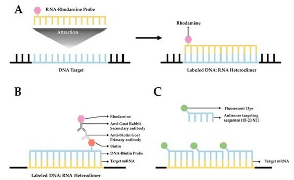 Fig. 1 A simplified diagram depicting FISH. (Tingey M, et al., 2022)
DNA FISH
DNA FISH is a powerful technique used for the visualization and mapping of specific DNA sequences within the genome. By employing fluorescent probes that hybridize to complementary DNA targets, DNA FISH enables the visualization of chromosomal abnormalities, gene copy number variations, and the spatial organization of genetic loci within the nucleus. This technique has been instrumental in cytogenetic analysis, cancer research, and prenatal diagnosis, offering valuable diagnostic and research applications.
RNA FISH
RNA FISH allows for the visualization and localization of specific RNA molecules within cells. By utilizing fluorescent probes that target RNA transcripts, researchers can study gene expression patterns, RNA localization, and transcriptional dynamics at the single-cell level. This technique has significantly contributed to our understanding of cellular processes such as mRNA localization, non-coding RNA dynamics, and viral RNA replication, making it a vital tool in molecular and cell biology research.
Comet-FISH
Comet-FISH, also known as single-cell gel electrophoresis, is a specialized FISH technique used to assess DNA damage and repair at the individual cell level. By combining the alkaline comet assay with FISH, researchers can visualize and quantify DNA breaks and chromosomal aberrations in individual cells, providing valuable insights into genotoxicity, mutagenesis, and DNA repair mechanisms. Comet-FISH has found applications in toxicology, environmental monitoring, and cancer research, enhancing our understanding of DNA damage at a single-cell resolution.
Flow-FISH
Flow-FISH combines FISH technology with flow cytometry, allowing for the simultaneous characterization and quantification of specific nucleic acid sequences in individual cells within a heterogeneous cell population. This technique enables high-throughput analysis of DNA or RNA targets, making it a valuable tool for studying cell cycle dynamics, gene expression heterogeneity, and identifying rare cell populations. Flow-FISH has been used extensively in cancer research, immunology, and microbial ecology to gain insights into cellular diversity and functional heterogeneity.
Immuno-FISH
Immuno-FISH is a technique that combines immunofluorescence staining with FISH to visualize specific proteins and nucleic acid sequences within cells simultaneously. This technique uses fluorescently labeled antibodies along with FISH probes to correlate the spatial distribution of proteins with the localization of DNA or RNA targets. It facilitates the study of protein-nucleic acid interactions, subcellular localization, and cellular signaling pathways. This technique has been crucial in revealing the spatial arrangement of genes, chromatin modifications, and protein-DNA interactions in various biological contexts, such as developmental biology and disease pathology.
Multiplex-FISH
Multiplex-FISH expands the capabilities of traditional FISH by allowing the simultaneous visualization of multiple DNA or RNA targets within the same sample. By utilizing a combination of differently labeled probes, researchers can generate complex multicolor FISH images, enabling the study of chromosomal rearrangements, gene expression networks, and spatial relationships between genomic loci. Multiplex-FISH has been invaluable in cancer cytogenetics, microbial ecology, and genomic profiling, offering a comprehensive view of genetic and transcriptomic landscapes within cells and tissues.
Multi-locus or ML-FISH
Multi-locus or ML-FISH is a specialized FISH technique designed to visualize and map multiple genetic loci or sequences within the genome. This technique allows for the simultaneous detection and localization of multiple genomic targets, offering insights into chromosomal organization, gene clustering, and genomic rearrangements. ML-FISH has been instrumental in studying genome architecture, chromosomal translocations, and gene regulation, providing a detailed understanding of nuclear organization and spatial relationships between genomic loci.
Quantitative FISH
This approach provides objective and quantitative measurements of FISH signals, allowing for the precise characterization of DNA or RNA targets in diverse biological samples. Quantitative FISH has emerged as a valuable tool for studying gene expression dynamics, chromosomal organization, and spatial relationships between genomic loci. By integrating advanced microscopy, image analysis, and computational techniques, researchers can extract quantitative data from FISH images, enabling the accurate assessment of gene copy numbers, RNA expression levels, and subcellular localization patterns.
Reverse FISH
Reverse FISH is a specialized FISH technique utilized for the identification and isolation of specific DNA sequences within complex genomes. This approach involves the hybridization of fluorescently labeled DNA probes to target sequences, followed by the retrieval and amplification of the hybridized DNA fragments. Reverse-FISH enables the targeted recovery and subsequent characterization of specific genomic regions, facilitating the study of gene structure, chromosomal organization, and the identification of novel DNA sequences. This technique has found applications in genomic mapping, DNA sequencing, and the isolation of genetic elements of interest, offering a valuable tool for the comprehensive analysis of complex genomes.
Creative Bioarray Relevant Recommendations
| Product/Service Types | Description |
| Fluorescent In Situ Hybridization (FISH) Service | Creative Bioarray offers a range of different FISH services including metaphase and interphase FISH, fibre-FISH, RNA-FISH, M-FISH, 3D-FISH, flow-FISH, FISH on paraffin sections, and immune-FISH. |
| FISH Probe Design, Synthesis, and Testing Service | Creative Bioarray is capable of developing custom FISH probes. Apart from that, we also offer mRNA ISH/FISH probes, miRNA ISH/FISH probes, and lncRNA ISH/FISH probes. |
| FISH Quality Control Services | Creative Bioarray has years of experience in performing FISH, and cytogenetic services, we can also provide custom FISH quality control service for your lab's needs! |
Reference
- Tingey M, et al. (2022). "Technologies Enabling Single-Molecule Super-Resolution Imaging of mRNA." Cells. 11 (19): 3079.