ImmunoFISH Analysis (FISH+IHC)
- Service Details
- Features
- FAQ
- Explore Other Options
ImmunoFISH Analysis (FISH+IHC) is Creative Bioarray's premium service in molecular and cellular biology, integrating the cutting-edge technologies of immunohistochemistry (IHC) and fluorescence in situ hybridization (FISH). This service empowers researchers to conduct precise analyses at the cellular microstructural and dynamic process levels, offering detailed insights into the DNA sequences and specific protein localizations and interactions within cells.
FISH technology employs fluorescent probes to bind with high specificity to particular DNA or RNA regions, aiding in the revelation of gene expression patterns and chromosomal structure abnormalities in both normal and diseased cells. On the other hand, IHC emphasizes the visualization of specific proteins through antigen-antibody interactions. Leveraging high-resolution fluorescence microscopy, ImmunoFISH quantifies single transcription and protein signals, enabling spatial and organizational analysis at the cellular level. This is exceptionally beneficial for uncovering the mechanisms of disease occurrence and developing diagnostic and therapeutic strategies. Thus, the combination of FISH and IHC offers a powerful and detailed spatial and functional analysis tool for cancer research, disease diagnosis, and precision medicine, holding immense potential for future applications.
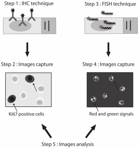 Fig. 1. Overview of ImmunoFish procedure (Céline Duval, Tayrac M D, et al., 2016).
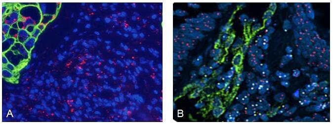 Fig. 2. (A) Lung cancer with human PPIB positive control mRNA probe (Red), CAM5.2 acidic cytokeratin (green) with DAPI as counterstain; (B) Human Chromosome X (Red), Y (Yellow) FISH and immunohistochemical stain using anti-von Willebrand factor (Green) on bone marrow transplant (Martínez-Ramírez A, Cigudosa JC, et al., 2004).
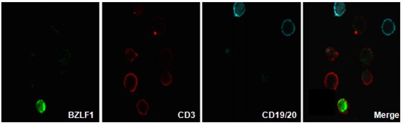 Fig. 3. Detection of EBV genomes in plasmablasts/plasma cells and non-B cells in the blood of most patients with EBV lymphoproliferative disorders using Immuno-FISH (Calattini S, Sereti I, et al., 2010).
Creative Bioarray's ImmunoFISH analysis service, backed by a professional technical team and state-of-the-art equipment, is dedicated to providing the most accurate and reliable analytical results for biomedical research.
Advantages of ImmunoFISH Technology
- Multiple labeling: Capable of observing the spatial relationship of multiple targets simultaneously.
- High specificity: Due to the highly specific binding between antigens and antibodies, the ImmunoFISH technique can accurately identify target molecules.
- High sensitivity: Fluorescent labeling allows clear observation of hybridization signals under a microscope, enabling highly sensitive detection of target molecules.
- Diverse applications: Beyond detecting chromosomal abnormalities and gene mutations, ImmunoFISH technology is applicable in studying biological processes such as protein-DNA interactions. Suitable for cancer research, molecular pathology studies, and drug development, among various other scenarios.
Our ImmunoFISH Analysis Service Include
- Sample preparation and preprocessing:
We accept a variety of tissue samples, including paraffin-embedded tissues, frozen sections, and cell culture samples.
Offers professional sample handling, including cell fixation and permeabilization, to ensure quality for experimental requirements. - FISH analysis:
Designs and prepares specific FISH probes based on target sequences.
Detects and visualizes target RNA/DNA sequences under a microscope for accurate localization. - IHC analysis:
Uses immunohistochemical staining methods to identify specific proteins or antigens. - Image acquisition:
Equipped with advanced microscopes and imaging systems to ensure high-quality image acquisition. - Data analysis and reporting:
Provides comprehensive data reports, including image processing and quantitative analysis, with clear visuals and graphs. - Consultation services:
Offers one-on-one expert consultation services to help clients understand results and provide recommendations for further research.
Our ImmunoFISH Analysis Service Advantages
- Expert team: Our team is composed of experts with years of experience in molecular biology and cytogenetics.
- Advanced equipment: Equipped with internationally leading microscopes, imaging systems, and image analysis software, supporting high-quality image acquisition and analysis.
- Strict quality control: Rigorously controlled quality standards from sample processing to data analysis ensure the reliability of experimental results.
- Rapid response and efficient service: Provides prompt customer service, ensuring that client needs are met in a timely manner.
Applications of ImmunoFISH Includes but Not Limited To
- Cancer diagnosis and research: ImmunoFISH can be used to analyze gene amplification and deletions in cancer tissues and identify cancer markers.
- Genetic disease diagnosis: Detects chromosomal aberrations in prenatal and neonatal diagnoses.
- Plant meiosis research: Frequently employed in studying the meiosis process in plants, particularly in cereal crops.
- Cell biology research: Useful in analyzing the location and changes of DNA and proteins during the cell cycle as well as chromatin structure within cells.
- Developmental biology research: Observes gene and protein expression patterns during embryonic development.
FAQ
1. What types of samples can be used for ImmunoFISH analysis?
We accept a variety of sample types, including paraffin-embedded tissues, frozen sections, and cell culture samples. Once received, the samples are optimized accordingly. Please consult our customer service team for specific sample requirements.
2. Is there a minimum sample requirement?
For cellular samples, we recommend at least 2 x 10⁶ cells per well. For tissue samples, at least 100 mg of fixed tissue is required.
3. What does the data report include?
We provide a comprehensive data report, including raw data, processed images, quantitative analysis results, and professional interpretation.
4. How long does ImmunoFISH analysis take?
From sample receipt, the turnaround time is typically 2-3 weeks; however, the specific time depends on the detection target and analysis complexity. We will maintain close communication with you throughout the process.
5. What types of molecules can ImmunoFISH detect?
Our ImmunoFISH service can simultaneously detect RNA/DNA sequences and various proteins.
Pricing and Ordering
Our customer service representatives are available 24 hours! Thank you for choosing Creative Bioarray for your ImmunoFISH analysis needs. For special inquiries or further assistance, please contact us or reach out online.
Explore Other Options