Chronic obstructive pulmonary disease (COPD) Models
Chronic obstructive pulmonary disease (COPD) is a leading cause of morbidity and mortality worldwide. It is characterized by chronic airway inflammation, excessive mucus secretion, airway remodeling, and emphysema, leading to decreased lung function and difficulty breathing. There is no effective treatment for COPD because the underlying mechanisms of COPD are poorly understood at the molecular level. However, animal experiments continue to treat all chronic diseases, including diseases that affect the airways and lungs.
Creative Bioarray provides various COPD models induced by Cigarette-smoke, elastase, and LPS, etc. We wholeheartedly provide our customers with assistance in drug development and mechanism exploration with the highest quality service.
COPD Models
- Cigarette smoke (CS)
Smoking is a significant risk factor for COPD and the most common COPD inducer in in vivo studies. In addition, for mainstream cigarette smoke, environmental cigarette smoke may also cause respiratory symptoms and COPD. CS has been proved to induce many features of COPD in animals, including pulmonary infiltration of macrophages and airway fibrosis, neutrophils, and emphysema.
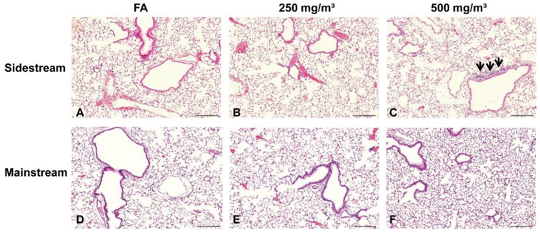 Figure 1. H&E staining of lung tissue slides from mice filter air (A and D); Sidestream (B and C) and mainstream (E and F) exposed to CS(John et al., 2013).
- Elastase
The currently accepted hypothesis that cigarette smoke induces emphysema is the protease-antiprotease hypothesis. Elastase is a proteolytic enzyme released by activated neutrophils in the lungs, causing alveolar tissue and emphysema rupture. The elastase model involves instilling elastase (such as porcine pancreatic elastase (PPE), human neutrophil elastase, and papain) in the lungs, which leads to tissue damage and the development of emphysema. This model is used to induce an inflammatory response to initiate and continue the inflammatory response seen in COPD.
 Figure 2. An example of a mouse emphysema model induced by elastase. The figure contrasts a representative control lung to a representative elastase-treated lung, with an overall increase in air space size of ~60%(Wright et al., 2008).
- Lipopolysaccharide (LPS)
LPS instillation can induce a short-term model of COPD with specific human characteristics of the disease. Animal chorionic exposure to LPS has been shown to induce the pathological features of COPD, such as lung inflammation and airway hyperresponsiveness (AHR), and structural changes in the lungs. LPS exposure twice a week induces an inflammatory response after 12 weeks. When administered alone or in combination with CS, LPS can induce acute exacerbations of COPD.
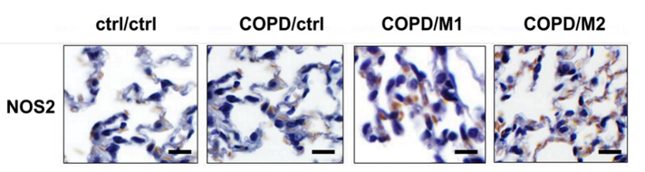 Figure 3. Examples of Immunohistochemical staining of lung tissues of ctrl, COPD, COPD-M1, and COPD-M2 groups at 2 hours post-injection of macrophages using iNOS(Faraj et al., 2014).
- Combined Inducers
Animal models that mimic different aspects of inflammatory responses in COPD could be developed by concomitant use of different inducers such as CS, LPS, and PPE. For example, mice could be intranasally challenged with PPE and LPS for 4 weeks to induce COPD-like lung inflammation.
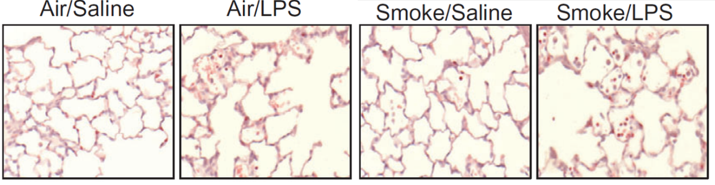 Figure 4. Neutrophils were assessed in the lung tissue by histological assessment(Hardaker et al., 2010).
Species
- Mouse
- Rat
Available Assays
- Cytotoxicity assays
- Measurement of cytokines
- Flow cytometric analysis of cell death
- Measurement of serum liver enzymes
- Hematoxylin and eosin staining (H&E)
- Immunohistochemistry
- Quantitative real-time PCR
- TUNEL stain
- Western blot analysis
- Gene expression analysis
Applications
- To evaluate the therapeutic and preventive effects of drugs on the occurrence and development of diseases.
- To explore the mechanism of drug action.
- To verify the function of new genes
Quotation and ordering
If you have any special needs or questions regarding our services, please feel free to contact us. We look forward to cooperating with you in the future.
References
- Faraj, A. A.; et al. Preferential Macrophage Recruitment and Polarization in LPS-Induced Animal Model for COPD: Noninvasive Tracking Using MRI. PLOS ONE, (2014), 9(3), e90829.
- Hardaker, E. L.; et al. Exposing rodents to a combination of tobacco smoke and lipopolysaccharide results in an exaggerated inflammatory response in the lung. British Journal of Pharmacology, (2010), 160(8), 1985-1996.
- John, G.; et al. The composition of cigarette smoke determines inflammatory cell recruitment to the lung in COPD mouse models. Clinical Science, (2013), 126(3), 207-221.
- Wright, J. L.; et al. Animal models of chronic obstructive pulmonary disease. American Journal of Physiology - Lung Cellular and Molecular Physiology, (2008), 295(1), L1-L15.