3D Co-culture Models Service
Cell cultures have played a significant role in the academy and pharmaceutical industry, but conventional 2D cultures fail to provide physiological relevance and communication network as compared to in vivo conditions, since they are unable to mimic the complexity of cellular microenvironment, an essential part for the cell behavior and systematic investigation. Using 2D cell culture systems can limit the predictive power of the cellular interactions. However, the 3D in vitro cultures provide a bridge between the conventional 2D cultures and in vivo studies. It was shown that in vitro 3D cultures recapture physiological cell-cell and cell-environment interactions (i.e., cellular morphology, cell differentiation, proliferation and gene expressions) with a greater relevant to animal and human studies, thus effectively facilitating the research progresses.
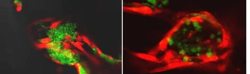
3D co-culture cell models now serve as important tools for solving scientific questions, which can be developed in different formats, depending on research goals and downstream usage. Co-culture cell models can emulate a variety of pathological conditions such as different stages of tumor invasion and metastasis, and identify of stromal components associated with pathological process.
Creative Bioarray 3D co-culture models service advantages
- Make your 3D co-culture models by using cells form our comprehensive human and animal cell bank or sending your own cells.
- Ready-to-use 3D cell cultures are highly functional and are amenable to high/live content imaging and a wide range of functional endpoint assays.
- Choose from numerous technologies (including scaffold-based and scaffold-free culture methods) to build your 3D co-culture models.
- Other 3D-based assays services are available at creative bioarray to fulfill your downstream research needs.
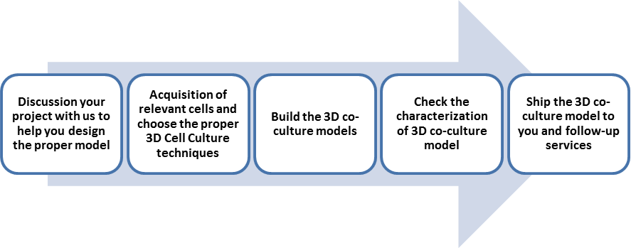
Workflow
Applications
Creative Bioarray provides services for co-culture tumor cells with other supportive stromal cells such as endothelial cells, fibroblast, immune cells and stem cells. Other 3D co-culture models are also available on request.
Study examples
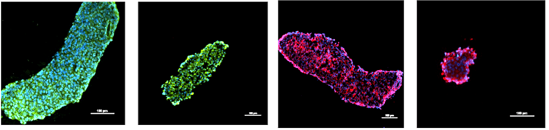
Figure. Representative images for different types of breast cancer cells co-cultured with Fibroblasts
Quotations and ordering
Our customer service representatives are available 24hr a day!
References
- Jaganathan, H., et al. Three-dimensional in vitro co-culture model of breast tumor using magnetic levitation. Scientific reports. 2014, 4.
- Sawant, S., et al. Establishment of 3D Co-Culture Models from Different Stages of Human Tongue Tumorigenesis: Utility in Understanding Neoplastic Progression. PloS one. 2016, 11.8: e0160615.
- Jiang, Q., et al. a Novel 3D Culture Model to Mimic Hematopoietic Niche for HSCs Differentiating to Megakaryocyte with Deproteined and Degreased Human Bone or β-TCP. Blood. 2014, 124.21: 4360-4360.
Explore Other Options