Four-vessel Occlusion (4-VO) Model
If you're interested in exploring new therapies and discovering the mechanism behind ischemic stroke, Creative Bioarray offers a cutting-edge four-vessel occlusion (4-VO) model. This model is designed to provide our clients with a comprehensive understanding of the underlying causes of ischemic stroke, which can be invaluable in developing new treatments and improving patient outcomes. With our 4-VO model, you will have access to the latest research and technology, as well as a team of experienced professionals who are dedicated to helping you achieve your goals. So why wait? Contact Creative Bioarray now to learn more about our 4-VO model and how it can benefit your research.
Four-vessel occlusion (4-VO) rat model has gained widespread use in the scientific community for exploring the underlying mechanisms of brain damage resulting from transient global ischemia, which mimics the pathological scenarios associated with cardiac arrest. This model is achieved by permanently coagulating the two vertebral arteries and temporarily ligating the two common carotid arteries, resulting in transient ischemia confined to the forebrain while the blood supply to the hindbrain remains intact. This innovative approach allows for the induction of severe yet reversible forebrain ischemia, leading to consistent neuronal damage in rats.
Our Four-vessel Occlusion (4-VO) Model
- Available Animal
Rat - Group Setting
- Sham group
- Model group
- Three dose groups of the test compound
- Endpoints
- Infarct volume assessment: TTC staining
- Histology analysis (brain tissue): H&E staining, Nissl staining
- Behavioral tests (e.g. motor function, cognition, social behavior)
- Other customized endpoints: available upon request
……
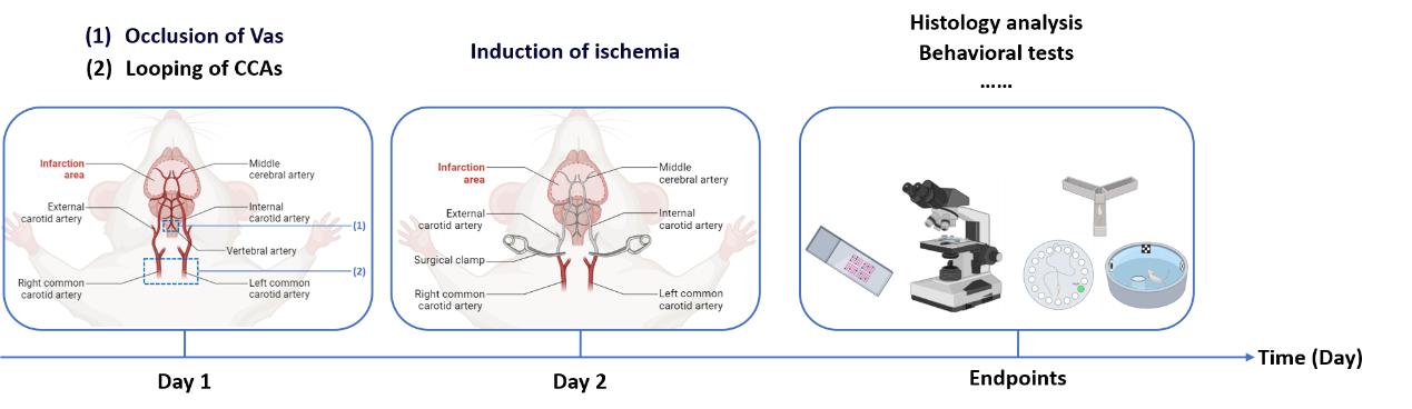 Fig. 1 Workflow of our 4-VO model
Example Data
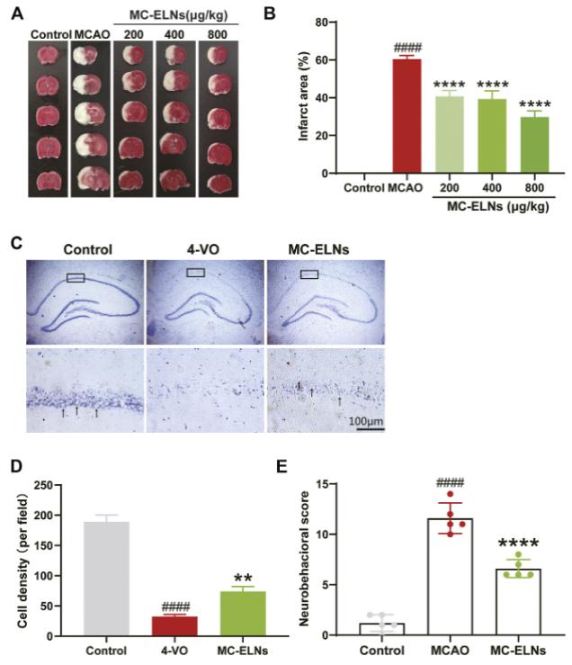 Fig. 2 MC-ELNs alleviate cerebral ischemia, promote hippocampus CA1 neuronal survival, and improve neurological function in transient cerebral ischemic rats. (A) Representative TTC staining images show the cerebral infarct area in MCAO rats treated with MC-ELNs of different concentrations. (B) Quantitative analysis of the percentage of the infarct area in each group. (C) Representative images show the cell density of hippocampus CA1 neurons by crystal violet staining. (D) Quantitative analysis of the number of neurons in the CA1 region of the hippocampus. (E) mNSS neurological severity scores showed that MC-ELN treatment could improve neurological deficits in MCAO ischemic rats.
Quotation and Ordering
If you are interested in our services, we invite you to contact us without hesitation to discuss your specific requirements. Together, we can craft a tailored study plan that precisely meets your needs and objectives.
Additionally, we provide other ischemic stroke animal models that you may be interested in:
References
- Kim, H., et al. Protocol for establishing a global ischemia model using a 4-vessel occlusion in rats. STAR protocols, 2023, 4(4): 102630.
- Cai, H., et al. Momordica charantia exosome-like nanoparticles exert neuroprotective effects against ischemic brain injury via inhibiting matrix metalloproteinase 9 and activating the AKT/GSK3β signaling pathway. Frontiers in Pharmacology, 2022, 13: 908830.