
KP-4
Cat.No.: CSC-C6816J
Species: Homo sapiens (Human)
Source: Ascites Metastasis
Morphology: Epithelial-Like
- Specification
- Background
- Scientific Data
- Q & A
- Customer Review
KP-4 is a well-characterized human pancreatic ductal adenocarcinoma (PDAC) cell line established in 1990 from the ascitic fluid of a 50-year-old Japanese male patient with metastatic pancreatic ductal cell carcinoma. The cells exhibit an adherent, epithelial-like morphology with characteristic cobblestone-like growth patterns. Karyotypically, KP-4 has a modal chromosome number of approximately 52-54 and carries a heterozygous KRAS G12D (c.35G>A) activating mutation, a hallmark oncogenic driver in PDAC. Additionally, the cell line displays loss of the tumor suppressor p53, contributing to enhanced cell survival and apoptosis resistance. KP-4 shows low E-cadherin expression and high vimentin expression, indicative of an epithelial-to-mesenchymal transition (EMT) phenotype critical for PDAC progression and metastatic dissemination.
A defining feature of KP-4 is its constitutive secretion of parathyroid hormone-related peptide (PTHrP), which renders it a valuable model for studying PTHrP regulation and its role in humoral hypercalcemia of malignancy and bone metastasis. The cell line exhibits high tumorigenicity upon transplantation into immunodeficient mice, with preferential metastatic dissemination to the liver and lungs. KP-4 expresses pancreatic cancer-associated markers including cytokeratins, epithelial membrane antigen (EMA), and the elevated cancer antigen CA19-9. The line also demonstrates high expression of matrix metalloproteinases (MMPs) that facilitate tumor invasion and metastasis.
KP-4 is widely utilized in preclinical studies investigating PDAC metastasis mechanisms-particularly liver tropism-as well as chemoresistance patterns and novel targeted therapies against KRAS or TGF-β pathways. The cell line serves as a robust platform for developing orthotopic PDAC mouse models and studying tumor-stroma interactions.
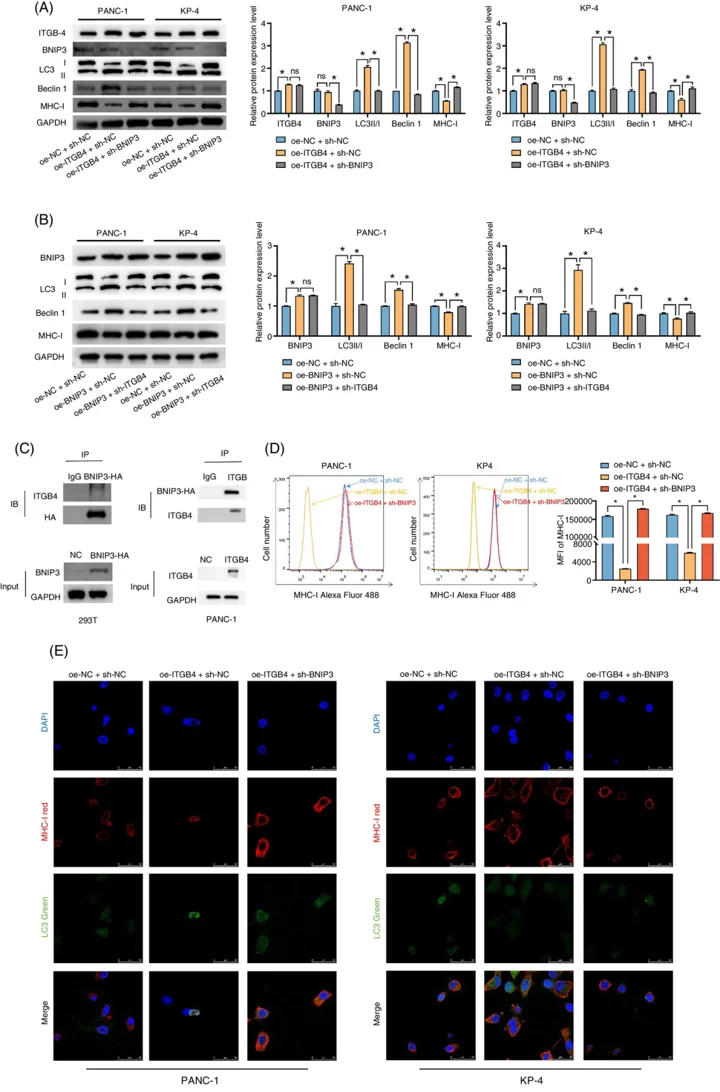
ITGB4/BNIP3 Activates Autophagy and Reduces MHC-I Expression to Mediate Tumor Immune Escape in Pancreatic Cancer Cell Lines
A certain connection between ITGB4 and cancer autophagy, making it a potential autophagic target. BNIP3 is a pro-apoptotic protein with a BH3 domain located on the mitochondrial membrane, associated with both cell apoptosis and autophagy. Therefore, this study aimed to explore the function mechanism of ITGB4 and BNIP3 in pancreatic cancer as well as their effects on cellular autophagy and MHC-I expression.
We transfected oe-ITGB4 and sh-BNIP3 into pancreatic cancer cell lines and detected the expression of BNIP3, MHC-I and autophagy-related proteins through WB. Autophagy was inhibited in the cells co-transfected with oe-ITGB4 and sh-BNIP3, and the expression level of MHC-I increased (Figure 1A). Furthermore, we conducted rescue experiments by knocking down ITGB4 in PANC-1 and KP-4 cells. WB results indicated that BNIP3 expression did not significantly change when ITGB4 was overexpressed or down-expressed, suggesting that ITGB4 did not directly influence BNIP3 expression (Figure 1B). Therefore, we assumed that cell autophagy was controlled by the mutual binding between ITGB4 and BNIP3. This hypothesis was verified through Co-IP experiments (Figure 1C). Furthermore, a comparable pattern in the MHC-I expression levels on the surface of pancreatic cancer cells under various treatments was revealed by flow cytometry (Figure 1D). Next, we observed the co-localization relationship between MHC-I and autophagosomes under confocal microscopy. As shown in Figure 1E, when cells were simultaneously transfected with oe-ITGB4 and sh-BNIP3, the degree of co-localization of MHC-I and LC3-labelled autophagosomes declined, which is consistent with the previously mentioned increasing trend of MHC-I expression.
Ask a Question
Write your own review
- You May Also Need

Description: Human moderately differentiated adenocarcinoma cell line established from liver metastasis.

Description: Established in 1985 from the primary ductal pancreatic adenocarcinoma (grade II) from a 44-year-old woman; reported as inducing metastasis in nude mice and as secreting proteinases (e.g. urokinase, ...

Description: established from the pancreas of a 81-year-old male patient with pancreatic ductal adenocarcinoma

Description: The MIA PaCa-2 cell line was established from tumor tissue of the pancreas obtained from a 65-year-old Caucasian male in 1975 by A. Yunis, et al. These cells are described to express human colony ...

Description: Established in 2010 from the pancreatic tumor of a 46-year old man with poorly differentiated ductal adenocarcinoma (high grade G3, pT3pN1)
Description: Established from the malignant ascites of a 66-year-old Japanese man with pancreas carcinoma in 1984
- Adipose Tissue-Derived Stem Cells
- Human Neurons
- Mouse Probe
- Whole Chromosome Painting Probes
- Hepatic Cells
- Renal Cells
- In Vitro ADME Kits
- Tissue Microarray
- Tissue Blocks
- Tissue Sections
- FFPE Cell Pellet
- Probe
- Centromere Probes
- Telomere Probes
- Satellite Enumeration Probes
- Subtelomere Specific Probes
- Bacterial Probes
- ISH/FISH Probes
- Exosome Isolation Kit
- Human Adult Stem Cells
- Mouse Stem Cells
- iPSCs
- Mouse Embryonic Stem Cells
- iPSC Differentiation Kits
- Mesenchymal Stem Cells
- Immortalized Human Cells
- Immortalized Murine Cells
- Cell Immortalization Kit
- Adipose Cells
- Cardiac Cells
- Dermal Cells
- Epidermal Cells
- Peripheral Blood Mononuclear Cells
- Umbilical Cord Cells
- Monkey Primary Cells
- Mouse Primary Cells
- Breast Tumor Cells
- Colorectal Tumor Cells
- Esophageal Tumor Cells
- Lung Tumor Cells
- Leukemia/Lymphoma/Myeloma Cells
- Ovarian Tumor Cells
- Pancreatic Tumor Cells
- Mouse Tumor Cells