Cat Tumor Cells
Cat (feline) tumor cells are essential models in veterinary oncology and comparative cancer research. Feline cancers, especially lymphomas and sarcomas, show interesting similarities with human cancers, and are thus useful for translational studies.
We maintain a selected panel of feline tumour cells for research on spontaneous and virus-associated cancers in cats, providing knowledge that bridges veterinary medicine and human oncology. These models are of great relevance to the study of retroviral pathogenesis as feline leukaemia virus (FeLV) is prevalent in cat populations.
Feline-Specific Veterinary Models Viral Oncology Translational Value

Key Features & Expertise
Specialized feline tumor models for focused research
Authentic Feline Cancer Models
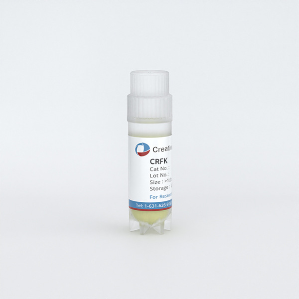
- Well-established feline cell lines including S+L-CAT2, CRFK, and PG-4
- Representative models for key feline cancer research areas
- Essential for veterinary oncology and comparative studies
FeLV-Related Research Tools
- Models for studying feline leukemia virus (FeLV) pathogenesis
- Tools for investigating retroviral transformation in cats
- Supports vaccine and therapeutic development
Reliable & Characterized
- Species-authenticated feline cell lines
- Quality-controlled for consistent performance
- Provided with optimized culture protocols
FAQ
What makes feline tumour cells so important for cancer research?
Feline cancers (especially lymphomas and injection-site sarcomas) are biologically very similar to human cancers. Spontaneous FeLV-associated lymphomas also occur in cats and provide a spontaneous model of retroviral carcinogenesis that is informative for veterinary and human medicine.
Can these cell lines be used for FeLV research?
Yes. S+L-CAT2 and other feline cell lines are of particular value for studies of FeLV, including virus propagation, neutralisation assays, and investigation of mechanisms of viral transformation. They are important tools for the development of diagnostics, vaccines and treatments for FeLV.
What are the primary applications of feline tumor cell lines?
Primary applications include: 1) Veterinary oncology drug research and development, 2) FeLV and feline retrovirus research, 3) Comparative oncology of feline and human cancer biology, 4) Virology and vaccine development, 5) Veterinary education.
How do I culture feline tumor cell lines?
Most feline cell lines grow well in standard media (DMEM or RPMI) containing foetal bovine serum at 37°C in the presence of 5% CO₂. Detailed protocols for each cell line are given. For example, the CRFK line is known to grow well and is commonly used for the isolation and propagation of feline viruses.
Filters Clear all filters
Species
- African clawed frog (1)
- American mink (1)
- Asian tiger mosquito (1)
- Atlantic salmon (1)
- Bluegill (2)
- Bluestriped grunt (1)
- Bovine (7)
- Brazilian free-tailed bat (1)
- Brown bullhead (2)
- Cabbage looper (1)
- Cabbage moth (6)
- Cat (3)
- Central mudminnow (1)
- Chicken (3)
- Chinese hamster (5)
- Chinook salmon (2)
- Chum salmon (1)
- Coho salmon (1)
- Common carp (2)
- Cotton-top tamarin (1)
- Dog (2)
- Fall armyworm (3)
- Fathead minnow (2)
- Fruit fly (1)
- Gilthead sea bream (2)
- Golden hamster (7)
- Goldfish (6)
- Gray dwarf hamster (1)
- Green monkey (2)
- Gypsy moth (1)
- Horse (1)
- Japanese eel (1)
- Japanese rice fish (7)
- Koi carp (1)
- Mouse (310)
- Mouse x Gray dwarf hamster (1)
- Mouse x Rat (20)
- Northern pike (1)
- Pig (3)
- Rabbit (2)
- Rainbow trout (3)
- Rat (114)
- Rhesus macaque (1)
- Salt marsh moth (1)
- Sheep (2)
- Snakehead murrel (2)
- Sockeye salmon (1)
- Vervet monkey (2)
- Zebrafish (2)
Source
- Abdomen (1)
- Adipose (2)
- Adrenal Gland (1)
- Aorta (4)
- Artery (1)
- Ascites (5)
- Ascites Metastasis (5)
- Bladder (11)
- Bladder Metastasis (1)
- Blastocyst (1)
- Blastula (1)
- Blood (7)
- Bone (6)
- Bone Marrow (14)
- Brain (24)
- Brain Metastasis (1)
- Breast (22)
- Caudal Peduncle (1)
- Caudal Trunk (2)
- Colon (6)
- Connective Tissue (7)
- Dermis (1)
- Embryo (29)
- Fetus (2)
- Fin (9)
- Glomerulus (2)
- Head Kidney (2)
- Heart (4)
- Hemolymph (1)
- Ileum (1)
- Intestine (9)
- Jejunum (1)
- Kidney (18)
- Liver (22)
- Lung (16)
- Lymph Node (2)
- Lymph Node Metastasis (1)
- Muscle (3)
- Ovary (8)
- Pancreas (9)
- Peripheral Blood (7)
- Peripheral Nervous System (21)
- Pituitary Gland (7)
- Prostate (3)
- Rectum (2)
- Skeletal Muscle (4)
- Skin (10)
- Small Intestine (3)
- Smooth Muscle (2)
- Soft Tissue (1)
- Spinal Cord (2)
- Testis (6)
- Thymus (5)
- Thyroid Gland (1)
- Trachea (1)
- Uterus (1)
Disease
- Bovine Leukemia (2)
- Canine Histiocytic Sarcoma (1)
- Chicken Bursal Lymphoma (2)
- Goldfish Erythrophoroma (4)
- Hamster Kidney Tumor (1)
- Hamster Pancreatic Ductal Adenocarcinoma (1)
- Hamster Uterine Leiomyosarcoma (1)
- Medaka Hepatoma (2)
- Mouse Bladder Transitional Cell Carcinoma (1)
- Mouse Chondrosarcoma (1)
- Mouse Colon Adenocarcinoma (3)
- Mouse Ependymoma (2)
- Mouse Erythroid Leukemia (13)
- Mouse Fibrosarcoma (5)
- Mouse Glioblastoma (1)
- Mouse Hemangioendothelioma (1)
- Mouse Hepatocellular Carcinoma (1)
- Mouse Insulinoma (3)
- Mouse Islet Cell Adenoma (1)
- Mouse Kidney Carcinoma (1)
- Mouse Leukemia (10)
- Mouse Leydig Cell Tumor (1)
- Mouse Lymphoma (8)
- Mouse Mammary Gland Malignant Neoplasm (21)
- Mouse Melanoma (9)
- Mouse Multiple Myeloma (5)
- Mouse Myeloid Leukemia (3)
- Mouse Neoplasm (1)
- Mouse Neuroblastoma (21)
- Mouse Oral Cavity Squamous Cell Carcinoma (1)
- Mouse Osteosarcoma (3)
- Mouse Pituitary Gland Neoplasm (1)
- Mouse Precursor T Cell Lymphoblastic Lymphoma/Leukemia (2)
- Mouse Pulmonary Adenoma (1)
- Mouse Pulmonary Malignant Tumor (3)
- Mouse Pulmonary Squamous Cell Carcinoma (1)
- Mouse Rectum Carcinoma (2)
- Mouse Reticulum Cell Sarcoma (2)
- Mouse Sarcoma (1)
- Mouse Teratocarcinoma (8)
- Mouse Thymic Lymphoma (3)
- Rat C-Cell Carcinoma (1)
- Rat Cholangiocarcinoma (1)
- Rat Colon Adenocarcinoma (5)
- Rat Digestive System Neoplasm (1)
- Rat Fibrosarcoma (1)
- Rat Hepatocellular Carcinoma (20)
- Rat Histiocytic Sarcoma (1)
- Rat Insulinoma (2)
- Rat Leukemia (1)
- Rat Leydig Cell Adenoma (1)
- Rat Lung Carcinoma (1)
- Rat Malignant Glioma (4)
- Rat Malignant Meningioma (1)
- Rat Malignant Oligodendroglioma (2)
- Rat Malignant Thymoma (3)
- Rat Mammary Gland Adenocarcinoma (10)
- Rat Neuroblastoma (3)
- Rat Osteosarcoma (2)
- Rat Pituitary Gland Neoplasm (6)
- Rat Prostate Adenocarcinoma (3)
- Rat Rhabdomyosarcoma (1)
- Rat Sarcoma (2)
- Rat Squamous Cell Carcinoma (1)
- Rat Urinary Bladder Transitional Cell Carcinoma (2)
- Rat Urinary System Neoplasm (6)

Description: Species: cat; Transformed by: murine sarcoma virus
Description: CRFK cells were isolated from the cortical portion of the kidneys of a 10-12 week old normal female ...

Description: PG-4 was derived at NIH in 1980 from G355 cells by transformation with Moloney Murine sarcoma virus ...