Middle Cerebral Artery Occlusion (MCAO) Model
If you are seeking to understand the pathogenesis of stroke and evaluate the efficacy of your compounds, Creative Bioarray's stable middle cerebral artery occlusion (MCAO) model can be of great help. With this model, you can get a better understanding of how stroke occurs and test the efficacy of your compounds on the MCAO model to find the best therapeutic solution. Our MCAO model is designed to provide accurate and reliable results, making it an ideal choice for researchers and scientists who are looking to study stroke and its underlying mechanisms.
Stroke is a leading cause of mortality and adult disability worldwide. Among the cerebral vessels affected by ischemic stroke, the middle cerebral artery (MCA) and its branches are frequently involved. As a result, the MCA occlusion (MCAO) technique has emerged as a widely accepted animal model for focal cerebral ischemia. This model closely mimics the pathological and physiological changes in human stroke, making it a valuable tool for preclinical research.
The induction of the MCAO model in rats involves the temporary occlusion of the common carotid artery (CCA), followed by the insertion of a suture into the internal carotid artery (ICA) or the transected external carotid artery (ECA). The suture is then advanced until it interrupts the blood supply to the MCA. The most commonly used durations of transient MCAO using a suture in rats are 60 minutes, 90 minutes, and 120 minutes.
Our Middle Cerebral Artery Occlusion (MCAO) Model
- Available Animal
Rat - Group Setting
- Sham group
- Model group
- Three dose groups of the test compound
- Endpoints
- Infarct volume assessment: TTC staining
- Histology analysis (brain tissue): H&E staining, Nissl staining
- Behavioral tests (e.g. motor function, cognition, social behavior)
- Other customized endpoints: available upon request
……
 Fig. 1 Workflow of our MCAO model
Example Data
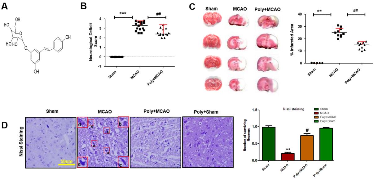 Fig. 2 Effect of polydatin on neurological scores, brain infarction and neurodegeneration. (A) Structure of polydatin. (B) Polydatin attenuated neurological deficits. Neurological test was conducted 24 h after ischemia. Poly+MCAO rats had significantly improvement of neurological deficits compared to MCAO group. (C) Brain coronal sections were stained with TTC, which distinguishes between ischemic and non-ischemic areas. (D) Representative photomicrograph of cresyl violet staining showing the extent of surviving neuron in the cortex. (a, b) Cytoplasmic swelling and scalloped neurons with intense cytoplasmic eosinophilia and nuclear basophilia. These changes result from neuronal ischemia, which impairs metabolism in the tissue. (c) Cytoplasmic fading of neurons, which invariably occurs in neurons at later stages of necrosis (ghost neurons). (d) Some cells had shrunken appearance along with pyknotic nuclei. Intensive neuropil vacuolation can be seen in ischemic cortex (MCAO).
Additionally, we provide other ischemic stroke animal models that you may be interested in:
Quotation and Ordering
With years of expertise in preclinical efficacy studies, Creative Bioarray stands as a trusted research partner, guiding you in selecting the most appropriate model for your specific research requirements. We pride ourselves on our ability to craft tailored study designs and protocols that align precisely with our clients' needs. For further insights into the aforementioned models or to discuss the possibility of introducing a novel model, please do not hesitate to contact us.
Reference
- Shah, F.A., et al. Polydatin attenuates neuronal loss via reducing neuroinflammation and oxidative stress in rat MCAO models. Frontiers in Pharmacology, 2019, 10: 663.