Inflammatory Bowel Disease (IBD) Models

Validated IBD Models
Chemically Induced Models
- Dextran Sulfate Sodium (DSS) induced model
- TNBS induced model
- Oxazolone induced model
- Indomethacin-induced colitis
Genetically Engineered Models
- IL-10 knockout colitis model
- Mdr1a knockout
- SAMP1/Fc mouse
Immune-Mediated Models

End-to-End IBD Research Services
Model Establishment
Validated induction protocols with high reproducibility
Customized Study Design
Strategies tailored to small molecules, biologics, and immunotherapies
Efficacy Evaluation
Clinical scoring (DAI score) and disease activity assessment
Mechanistic Analysis
Immune, molecular, and pharmacodynamic profiling
Chronic & Translational Studies
Long-term monitoring aligned with clinical relevance
Integrated Readouts
Histology, immune cell profiling, cytokine profiling, gene and protein expression analysis and optional RNA-seq.
Understanding Inflammatory Bowel Disease (IBD)
Inflammatory Bowel Disease (IBD) is a chronic, relapsing inflammatory disorder of the gastrointestinal tract, primarily encompassing Ulcerative Colitis (UC) and Crohn's Disease (CD). The global incidence of IBD continues to rise, creating substantial clinical and economic burdens worldwide.
Although both conditions involve intestinal inflammation, they differ significantly in anatomical distribution, immune mechanisms, and pathological progression—factors that critically influence preclinical model selection.
UC vs CD Comparison
| Feature | Ulcerative Colitis (UC)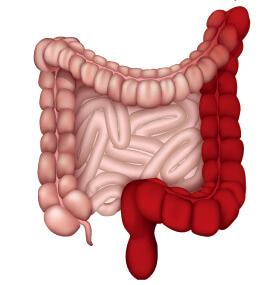
|
Crohn's Disease (CD)
|
|---|---|---|
| Distribution Pattern | Continuous inflammation in the colon | Segmental ("skip") lesions throughout the GI tract |
| Anatomical Location | Limited to the colon | Can affect any part of the GI tract (commonly ileum + colon) |
| Depth of Inflammation | Mucosal and submucosal layers | Transmural (full-thickness involvement) |
| Key Pathophysiology | Epithelial barrier disruption, neutrophil infiltration, ulceration | Granuloma formation, fibrosis, strictures, fistula development |
| Immune Profile | Th2-skewed cytokine response | Th1/Th17-dominant cytokine response |
| Common Complications | Severe bleeding, toxic megacolon | Strictures, abscesses, perianal disease |
The Challenge in IBD Drug Development
Despite advances in biologics targeting TNF-α, integrins, and IL-12/23 pathways, many patients experience incomplete response or disease relapse.
Major challenges include:
- Disease heterogeneity
- Limited predictive biomarkers
- Poor translational consistency between models and clinical outcomes
Given the complexity and heterogeneity of IBD, the choice of preclinical model directly impacts the predictability of therapeutic outcomes. At Creative Bioarray, we offer a comprehensive portfolio of validated IBD models designed to deliver robust, translational, and decision-ready data.
IBD Model Comparison
| Model | Advantages | Limitations | Suitable Applications | Clinically Relevant | Mechanism |
|---|---|---|---|---|---|
| DSS-induced colitis | Simple, reproducible, rapid | Innate immunity-dominated | Anti-inflammatory drugs, barrier repair agents | Ulcerative Colitis | Epithelial barrier disruption allows luminal antigens to trigger acute inflammation |
| TNBS-induced colitis | Crohn's-like Th1 response | Technically sensitive, variable | Immunomodulators, biologics | Crohn's Disease | Hapten-induced immune activation drives Th1 cytokine-mediated colitis |
| Oxazolone-induced colitis | Th2-driven, UC-like | Superficial mucosal damage | Biologics targeting Th2 pathways | Ulcerative Colitis | NKT cell activation induces epithelial damage and Th2 inflammation |
| IL-10 knockout | Spontaneous chronic colitis | Long modeling time | Mechanistic studies, long-term efficacy | Crohn's Disease | Loss of IL-10 regulation leads to uncontrolled Th1/Th17 immune responses |
| Adoptive T cell transfer | Chronic immune-driven colitis | Technically demanding | Biologics, immunotherapies | UC & Crohn's Disease | Activated T cells induce sustained intestinal inflammation (Th1/Th17) |
| Anti-CD40 antibody | Rapid immune activation | Acute inflammation, higher cost | Immune checkpoint modulators | Ulcerative Colitis | CD40-CD40L signaling activates innate immune cells (DCs/macrophages) |
Why Choose Creative Bioarray's IBD Models
Extensive Model Portfolio
Acute, chronic, genetic, and immune-mediated models covering all IBD research stages.
Technical Expertise
SOP-standardized procedures ensuring reproducible and high-quality data.
Customizable Study Design
Protocols tailored to your compound type and development goals.
Flexible and Integrated Endpoints
Clinical, histological, molecular, and immune biomarkers in one unified platform.
Partner With Us
Need guidance selecting the optimal IBD model for your therapeutic program?
Our experienced scientific team is ready to support your project from study design to data interpretation.
Talk to an Expert