Histological Staining Techniques: From Traditional Chemical Staining to Immunohistochemistry
Staining is a crucial step in histology to enhance the visibility of the fine structures of tissues. It increases the contrast of cells and their components, making them clearer for observation and analysis under a microscope.
Chemical Saining
Chemical staining is a traditional method that utilizes chemical dyes to color tissue sections. These dyes interact selectively with particular chemical components found in tissue which enables the visualization of different structures through distinctive colors and contrasts when observed under a microscope.
- HE staining
The Hematoxylin and Eosin (HE) staining technique stands as a fundamental approach within pathology research. Basic dye hematoxylin causes acidic components like nucleic acids in the nucleus and cytoplasm to develop blue-purple stains. The acidic dye Eosin colors basic proteins pink throughout the cytoplasm and extracellular matrix. The staining method enables researchers to distinguish the nucleus from cytoplasm while revealing diseased tissue markers and transformations with clarity.
Application: HE staining serves as a fundamental technique in histology and pathology as well as embryology to analyze tissue structures and disease states including apoptosis necrosis inflammation and tumor development.
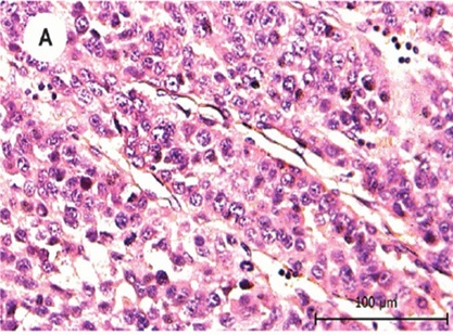 Fig. 1. Hematoxylin and eosin staining of the patient's primary tumor (Xu XL, Xing BC, et al., 2010).
- Masson's trichrome staining
Masson's Trichrome staining serves as a traditional technique for examining connective tissue. The staining method applies three different dyes namely iron hematoxylin for collagen fibers, fuchsin for muscle fibers and methylene blue for cytoplasm to achieve distinct coloration. Collagen takes on a blue color while muscle fibers display a red hue and cytoplasm shows up as black or dark gray.
Application: It is mainly used to observe the distribution and ratio of collagen and muscle fibers, commonly applied in studies of fibrous tissue proliferation, fibrosis, and the diagnosis of diseases such as cirrhosis and nephritis.
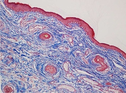 Fig. 2. Masson trichrome staining of eyelid specimens (Kocer AM, Sen EM, et al., 2022).
- Sirius red staining
Sirius red functions as an intense acidic anionic dye that possesses numerous sulfonic groups which attach to basic amino acids within collagen structures. Stained collagen fibers show red coloration and demonstrate birefringence which aids in differentiating collagen types.
Application: It is used to assess iron overload pathology and liver diseases and to detect the degree of fibrosis.
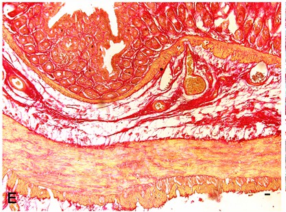 Fig. 3. Sirius red staining of collagen fibers of full-thickness normal rat colon (Segnani C, Ippolito C,et al., 2015).
- PAS glycogen staining
The PAS staining method uses oxidation of carbohydrates to create aldehyde groups that react with Schiff reagent to produce a purple color. This technique targets glycogen alongside polysaccharides and various mucopolysaccharides.
Application: This method serves as a common diagnostic tool for diabetes and myocardial lesions while also helping to identify tumors and research abnormal glucose metabolism and liver diseases.
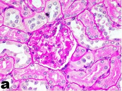 Fig. 4. Periodic acid–Schiff staining of kidney tissue (ElMosbah DE, Khattab MS, et al., 2022).
Table. 1. Other chemical staining method
| Staining Method | Staining Site | Common Staining Objects | Staining Result |
| TTC | Cytoplasm | Cells, tissues | Pink |
| Nissl Staining | Neuronal nuclei | Nervous tissue | Blue |
| Luxol Fast Blue (LFB) | Collagen fibers, reticular fibers | Connective tissue | Blue green |
| Golgi Staining | Neuronal process | Nerve cells | Black |
| Congo Red Staining | Collagen fibers, amyloid substances | Connective tissue | Red |
| Ferric Iron Blue (FJB) Staining | Elastic fibers | Connective tissue | Black |
| Eosinophilic Vascular Grafting (EVG) Staining | Collagen fibers, reticular fibers, elastic fibers | Connective tissue | Green |
| Oil Red O Staining | Neutral fat droplets | Adipose tissue | Orange-red |
| Ice-cut Oil Red O Staining | Neutral fat droplets | Adipose tissue | Orange-red |
| Sirius Red Staining | Collagen fibers | Connective tissue | Red |
| Periodic Acid-Schiff (PAS) Staining | Glycogen, polysaccharides | Liver, lung, etc. | Red |
| PAS with Silver Methenamine (PASM) Staining | Glycogen, polysaccharides | Liver, lung, etc. | Red |
| Weigert's Elastic Fiber Staining | Elastic fibers | Connective tissue | Black |
| Alcian Blue (AB) Staining | Chondroitin sulfate and mucopolysaccharide | Cartilage tissue | Blue |
| Mast Cell Staining | Mast cells | Mast cells | Red |
| Silver Staining | Reticular fibers | Connective tissue | Black |
| Methylene Blue Staining | Nuclear bodies, Nissl bodies | Nervous tissue | Blue |
| Goldner's Trichrome Staining | Skeletal muscle fibers | Skeletal muscle | Multiple colors |
Immunohistochemical (IHC)
Immunohistochemistry uses antibodies to find specific antigens (proteins) in tissues. With enzyme or fluorescent labels, it's possible to directly observe expression and distribution of particular proteins. IHC is categorized into immunofluorescence, enzyme-linked immunohistochemistry, immunoferrography, immunogold labeling, autoradiography, etc., according to the markers used.
- Immunofluorescence (IF)
Immunofluorescence: Immune antibodies can be marked with fluorescent dyes to visualize target antigens. In this approach, antibodies are labelled directly or indirectly with fluorescent dyes. When excited by light of a particular wavelength, these dyes emit visible light that signals where the antigen target is located. It is commonly used for intracellular studies and protein localization and it is applicable for multiplex experiments.
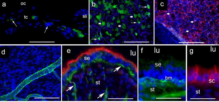 Fig. 5 Immunofluorescence (IF) staining in frozen sections (Lopez IA, Ishiyama G, et al., 2016).
- Enzyme-linked immunohistochemistry
Enzyme-linked immunohistochemistry shows the enzymatic association of antigen with antibody. Secondary antibodies are usually paired with enzymes (like horseradish peroxidase, HRP or alkaline phosphatase, AP), and then the substrates are added to do a colorimetric reaction. It is the technique most commonly used in pathology and diagnostics to show particular antigens in tissue sections.
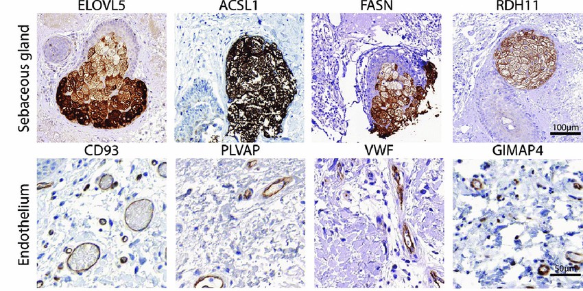 Fig. 6 Examples of immunohistochemical staining of proteins encoded by SkinSig marker genes (Shih BB, Nirmal AJ, et al., 2016).
- Immunogold labeling
Immunogold labels antibodies using gold particles, which are seen in an electron microscope. Gold atoms are dense black spots in electron micrographs that pinpoint the antigens of interest. This is mainly used to examine ultrastructures, and especially for high-resolution localization in electron microscopy.
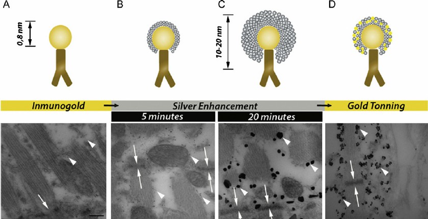 Fig. 7 Diagram of the immunogold labeling technique (Sirerol-Piquer MS, Cebrián-Silla A, et al., 2012).
Creative Bioarray Relevant Recommendations
| Products & Services | Description |
| Special Staining Services | Creative Bioarray can offer a variety of comprehensive and reliable special staining for sectioned paraffin embedded tissues or cells to fit all individual needs. |
| Immunohistochemistry (IHC), Immunofluorescence (IF) Service | Creative Bioarray offers the best immunohistochemistry and immunofluorescence service. |
References
- Xu XL, Xing BC, et al. The properties of tumor-initiating cells from a hepatocellular carcinoma patient's primary and recurrent tumor. Carcinogenesis. 2010. 31(2):167-74.
- ElMosbah DE, Khattab MS, et al. The anti-inflammatory effect of myrrh ethanolic extract in comparison with prednisolone on an autoimmune disease rat model induced by silicate. Inflammopharmacology. 2022. 30(6):2537-2546.
- Kocer AM, Sen EM, et al. The histopathological findings in excised upper eyelids of patients with dermatochalasis following collagen cross-linking treatment. Graefes Arch Clin Exp Ophthalmol. 2022. 260(8):2737-2743.
- Lopez IA, Ishiyama G, et al. Immunohistochemical techniques for the human inner ear. Histochem Cell Biol. 2016. 146(4):367-87.
- Shih BB, Nirmal AJ, et al. Derivation of marker gene signatures from human skin and their use in the interpretation of the transcriptional changes associated with dermatological disorders. J Pathol. 2017. 241(5):600-613.
- Sirerol-Piquer MS, Cebrián-Silla A, et al. GFP immunogold staining, from light to electron microscopy, in mammalian cells. Micron. 2012. 43(5):589-99.
- Segnani C, Ippolito C, et al. Histochemical Detection of Collagen Fibers by Sirius Red/Fast Green Is More Sensitive than van Gieson or Sirius Red Alone in Normal and Inflamed Rat Colon. PLoS One. 2015. 10(12):e0144630.