
Ocular Cells
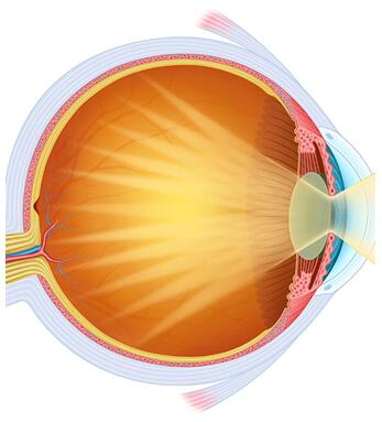

Ocular cells are highly specialized populations that maintain visual function, ocular barrier integrity, immune regulation, and retinal homeostasis. The eye contains diverse epithelial, endothelial, neuronal, stromal, and vascular-associated cell types that work together to support vision and protect delicate ocular tissues from injury and inflammation.
Our ocular cell collection provides physiologically relevant in vitro models for ophthalmology research, retinal vascular biology, neurodegeneration studies, ocular inflammation, glaucoma research, and drug discovery applications. The portfolio includes corneal, conjunctival, retinal, choroidal, trabecular meshwork, and retinal microvascular cell types, as well as ocular-supporting neuronal and immune-associated cells.

15+
Ocular Cell
Models

100 %
Mycoplasma-Free
Tested

24-72 h
Rapid
Delivery
Filter By:
Our Ocular Cell Portfolio Highlights

Comprehensive Ocular Cell Resources
Our collection covers major cell populations involved in ocular development, visual function, retinal homeostasis, and eye disease pathogenesis.
-
Anterior Segment & Ocular Surface Cells
- Conjunctival epithelial cells and fibroblasts
- Corneal endothelial cells and keratocytes
- Iris, lens, and ciliary epithelial cells
-
Retinal & Vascular Cell Models
- Retinal microvascular endothelial cells and pericytes
- Retinal pigment epithelial cells
- Choroidal fibroblasts and ocular vascular models
-
Advanced Reporter-Labeled Models
- GFP- and RFP-expressing retinal endothelial cells
- Suitable for live-cell imaging and angiogenesis studies
-
Flexible Research Solutions
- Ready-to-use cryopreserved primary cells
- Customized sourcing and project support available

Ophthalmology Research Applications
Our ocular cell models support studies of retinal disorders, glaucoma, ocular inflammation, corneal biology, and therapeutic development.
-
Retinal Disease Research
Retinal pigment epithelial cells, retinal endothelial cells, and pericytes are widely used in studies of diabetic retinopathy, retinal degeneration, and age-related macular degeneration.
-
Blood-Retinal Barrier Studies
Retinal vascular endothelial cells and pericytes provide valuable tools for investigating vascular permeability, angiogenesis, and neurovascular interactions.
-
Glaucoma & Anterior Segment Research
Trabecular meshwork cells and anterior segment epithelial cells support investigations into aqueous humor dynamics and glaucoma pathogenesis.
-
Ocular Drug Discovery
Primary ocular cells facilitate efficacy testing, toxicity assessment, target validation, and translational ophthalmic research.
What types of ocular cells are available?
Our portfolio includes conjunctival, corneal, retinal, choroidal, iris, lens, ciliary body, trabecular meshwork, vascular endothelial, pericyte, and retinal pigment epithelial cells representing major structures of the human eye.
Do you provide retinal disease research models?
Yes. We offer retinal pigment epithelial cells, retinal microvascular endothelial cells, retinal pericytes, and neuronal models commonly used for studies of diabetic retinopathy, retinal degeneration, and age-related macular degeneration.
Are reporter-labeled ocular vascular cells available?
Yes. GFP- and RFP-expressing retinal microvascular endothelial cells are available for angiogenesis research, cell tracking, live-cell imaging, and vascular biology studies.
What applications are supported by trabecular meshwork cells?
Trabecular meshwork cells are widely used in glaucoma research, aqueous humor outflow studies, intraocular pressure regulation investigations, and ophthalmic drug development.
How are ocular cells shipped and stored?
Cells are generally shipped cryopreserved on dry ice and should be stored in liquid nitrogen upon arrival. Detailed recovery and culture protocols are provided to support optimal viability and experimental reproducibility.