THP-1 l
Cat.No.: CSC-6320W
Species: Homo sapiens (Human)
Source: Blood; Peripheral Blood
Morphology: continuous culture, grown in suspension, morphology large, round, single cells
Culture Properties: suspension
- Specification
- Background
- Scientific Data
- Q & A
- Customer Review
Tissue: peripheral blood;
Tumor: leukemia, acute monocytic;
Derived from: THP-1
vWA: 16;
FGA: 24,25;
TH01: 8,9.3;
D18S51: 13,14;
D21S11: 30,31.2;
D8S1179: 10,14
The THP-1 cell line remains the global gold standard for modeling human monocytic and macrophage biology. Our enhanced THP-1 (THP-1-l) variant is specifically curated through strict lineage maintenance and low-passage expansion, ensuring the retention of robust phagocytic capacity, migratory responsiveness, and sensitivity to pro-inflammatory stimuli-features often lost in standard laboratory-maintained stocks.
- Optimized Differentiation Fidelity: THP-1-l exhibits superior sensitivity to PMA-induced differentiation, rapidly transitioning into a functional macrophage-like state. This cell line consistently generates homogeneous populations, which is essential for studying M1/M2 polarization and phenotypic plasticity.
- Predictable Inflammasome Kinetics: Characterized by high-fidelity activation of the NLRP3 inflammasome, this line provides a highly reliable platform for assessing cytokine release (e.g., IL-1β, TNF-α) and testing the inhibitory efficacy of novel anti-inflammatory compounds.
- Genetic Stability & Transduction Efficiency: By limiting passage numbers, we ensure higher structural integrity of the genome and reliable expression of cell surface markers (e.g., CD14, CD11b). This makes our THP-1-l the ideal substrate for CRISPR/Cas9 editing and lentiviral transduction to study host-pathogen interactions.
- High-Throughput Screening (HTS) Consistency: With consistent growth kinetics and standardized doubling times, our THP-1-l minimizes experimental variance, allowing for reproducible data collection across multi-well platforms for drug discovery and toxicity screening.
By choosing our low-passage THP-1-l, you invest in a highly validated, stable, and functionally superior model that brings unparalleled accuracy to your inflammatory and immunological research.
Platelet-Derived Microvesicles Modulate Cytokine and Lipid Mediator Profiles in THP-1 Monocytes
Monocytes are circulating immune cells that migrate to inflamed tissues and differentiate into macrophages, where they play a dual role in regulating pro-inflammatory and pro-resolving responses through cytokine and lipid mediator secretion. Platelet-derived microvesicles (PMVs), released during platelet activation, infiltrate inflamed areas and interact with monocytes and macrophages, facilitating the transfer of bioactive contents. While these interactions have been observed, their functional consequences on monocyte/macrophage inflammatory profiles remain poorly understood.
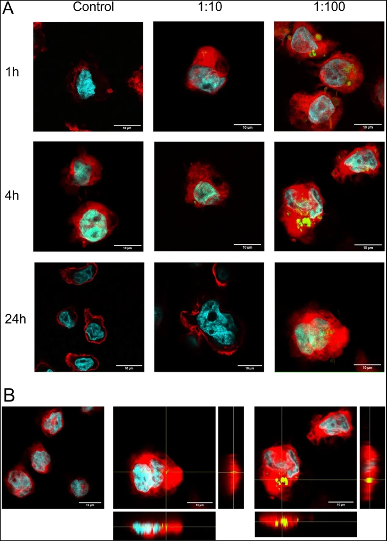
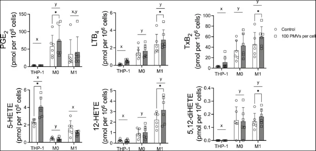
In this study, PMVs are shown to be internalized by human THP-1 monocytes. The interaction with THP-1 cells occurs rapidly, with 60 % of cells interacting with PMVs within one hour. When cells are differentiated to M0 and M1 macrophages, interactions with PMVs only peak after 24 h. Interaction of cells with PMVs resulted in an increased capacity to synthesize cyclooxygenase- and lipoxygenase-derived lipid mediators of inflammation, especially in M1 cells. Cytokine production was also influenced in a cell-state-dependent manner. PMVs had no impact on undifferentiated THP-1 cells but enhanced the production of several cytokines in M0 cells as well as IL-23 and IL-6 in M1 macrophages. When stimulated with lipopolysaccharides, PMV-treated M0 macrophages demonstrated elevated production of the anti-inflammatory cytokine IL-10, while M1 macrophages exhibited increased secretion of IL-1β, MCP-1, and IL-6, highlighting an effect on pro-inflammatory cytokine production. These findings reveal that PMVs selectively modulate the inflammatory cytokine and lipid mediator profiles of monocytes and macrophages depending on their differentiation state.
Ask a Question
Write your own review
- You May Also Need

Description: Established in 2007 from the bone marrow mononuclear cells of an 82-year-old Japanese man with diffuse large B-cell lymphoma in the leukemic phase

Description: Established from the bone marrow of a 28-year-old man who developed the terminal leukemic phase of lymphosarcoma in 1976

Description: This cell line was derived from the bone marrow aspirate of a 59 year old male with erythroleukemia that became acute myelogenous leukaemia.The cells form colonies in soft-agar in the presence of ...

Description: Established from the pleural effusion of a 24-year-old woman with recurrent anaplastic large cell lymphoma (ALCL); cells were described to clonally derive from T-lineage lymphoid cells and to be ...

Description: Established from a 37-year-old man at second (refractory/terminal) relapse of Hodgkin lymphoma (nodular sclerosing -> lymphocyte depleted/stage IIISA -> stage IV) after both combined chemo- and ...

Description: Established from the peripheral blood of a 10-year-old Caucasian boy with acute lymphoblastic leukemia (pre B-ALL) at diagnosis in 1993
- Adipose Tissue-Derived Stem Cells
- Human Neurons
- Mouse Probe
- Whole Chromosome Painting Probes
- Hepatic Cells
- Renal Cells
- In Vitro ADME Kits
- Tissue Microarray
- Tissue Blocks
- Tissue Sections
- FFPE Cell Pellet
- Probe
- Centromere Probes
- Telomere Probes
- Satellite Enumeration Probes
- Subtelomere Specific Probes
- Bacterial Probes
- ISH/FISH Probes
- Exosome Isolation Kit
- Human Adult Stem Cells
- Mouse Stem Cells
- iPSCs
- Mouse Embryonic Stem Cells
- iPSC Differentiation Kits
- Mesenchymal Stem Cells
- Immortalized Human Cells
- Immortalized Murine Cells
- Cell Immortalization Kit
- Adipose Cells
- Cardiac Cells
- Dermal Cells
- Epidermal Cells
- Peripheral Blood Mononuclear Cells
- Umbilical Cord Cells
- Monkey Primary Cells
- Mouse Primary Cells
- Breast Tumor Cells
- Colorectal Tumor Cells
- Esophageal Tumor Cells
- Lung Tumor Cells
- Leukemia/Lymphoma/Myeloma Cells
- Ovarian Tumor Cells
- Pancreatic Tumor Cells
- Mouse Tumor Cells