Atherosclerosis Model
Atherosclerotic cardiovascular disorder is increasingly the main cause of mortality worldwide. Atherosclerosis is characterized by arterial plaque and ultimately lead to acute myocardial infarction or stroke. Animal models of atherosclerosis are based on cholesterol-rich diet and manipulation of special genes. At present, mouse and rabbit models are commonly used in mechanism research and translational research. These models have made some contributions to the treatment and diagnosis of atherosclerotic disease.
Creative Bioarray focuses on drug research and development services and helps customers evaluate the drug efficacy and study the associated pathological mechanisms of atherosclerosis by atherosclerosis model.
Models available
- Hgih-fat diet induced APOE(−/−) mouse model
- High-Fat Diet-Induced Atherosclerosis Model
- Ballon Injury and High-Fat Diet-Induced Atherosclerosis Model
Species available
- Rabbit
- Mouse
- Rat
Our capabilities
- We screen novel test compounds targeting atherosclerosis.
- We use HE staining, Oil Red O and Masson trichrome staining for determination of atherosclerotic lesion.
Assays available
- Histopathological evaluation
- Cytokine analysis
With extensive experience in the field of cardiovascular disorder, we are confident to help you to overcome any upcoming challenges. Our experts are fully capable of customizing our protocols and assays to meet your specific needs. With our help, we wish to facilitate your research with high efficiency.
Study examples
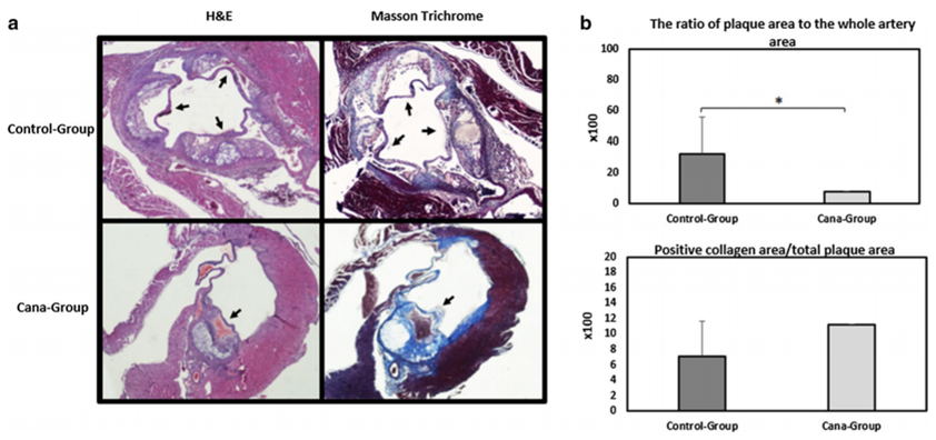 Figure. 1. Atherosclerotic plaque extension among APOE(−/−) mice on western diet treated with canaglifozin (Cana-group) or vehicle (control-group). a Selected 4 μm section images from the aortic root stained with H&E and Masson trichrome. Formation of atherosclerotic plaque was examined using H&E staining while histological examination of atherosclerotic plaque collagen content was assessed using Masson trichrome staining. b Quantifcation of plaque area is shown as a percentage of lumina stenosis by thickened intima. Collagen content was measured using quantifcation of Masson trichrome positive area over complete plaque area.
Quotation and ordering
If you have any special needs or questions regarding our services, please feel free to contact us. We look forward to cooperating with you in the future.
Reference
Nasiri-Ansari Νarjes, et al. Canagliflozin attenuates the progression of atherosclerosis and inflammation process in APOE knockout mice[J]. Cardiovascular Diabetology, 2018, 17(1):106.
For research use only. Not for any other purpose.
Disease Models
- Oncology Models
-
Inflammation & Autoimmune Disease Models
- Rheumatoid Arthritis Models
- Glomerulonephritis Models
- Multiple Sclerosis (MS) Models
- Ocular Inflammation Models
- Sjögren's Syndrome Model
- LPS-induced Acute Lung Injury Model
- Peritonitis Models
- Passive Cutaneous Anaphylaxis Model
- Delayed-Type Hypersensitivity (DTH) Models
- Inflammatory Bowel Disease Models
- Systemic Lupus Erythematosus Animal Models
- Oral Mucositis Model
- Asthma Model
- Sepsis Model
- Psoriasis Model
- Atopic Dermatitis (AD) Model
- Scleroderma Model
- Gouty Arthritis Model
- Carrageenan-Induced Air Pouch Synovitis Model
- Carrageenan-Induced Paw Edema Model
- Experimental Autoimmune Myasthenia Gravis (EAMG) Model
- Graft-versus-host Disease (GvHD) Models
-
Cardiovascular Disease Models
- Surgical Models
- Animal Models of Hypertension
- Venous Thrombosis Model
- Atherosclerosis model
- Cardiac Arrhythmia Model
- Hyperlipoidemia Model
- Doxorubicin-induced Heart Failure Model
- Isoproterenol-induced Heart Failure Model
- Arterial Thrombosis Model
- Pulmonary Arterial Hypertension (PAH) Models
- Heart Failure with Preserved Ejection Fraction (HFpEF) Model
- Cardio-Renal-Metabolic (CKM) Syndrome Model
-
Neurological Disease Models
- Alzheimer's Disease Modeling and Assays
- Ischemic Stroke Models
- Acute Spinal Cord Injury (ASCI) Model
- Traumatic Brain Injury (TBI) Model
- Hypoxic-Ischemic Encephalopathy (HIE) Model
- Tourette Syndrome (TS) Model
- Amyotrophic Lateral Sclerosis (ALS) Model
- Huntington's Disease (HD) Model
- Intracerebral hemorrhage (ICH) Models
- Schizophrenia Model
- Depression Models
- Pain Models
-
Metabolic Disease Models
- Type 1 Diabetes Mellitus Model
- Type 2 Diabetes Mellitus Model
- Animal Model of Hyperuricemia
-
Nonalcoholic Fatty Liver Disease Model
- High-Fat Diet-Induced Nonalcoholic Fatty Liver Disease (NAFLD) Model
- Methionine and Choline Deficient (MCD) Diet-Induced Nonalcoholic Fatty Liver Disease (NAFLD) Model
- Gubra-Amylin NASH (GAN) Diet-Induced Nonalcoholic Fatty Liver Disease (NAFLD) Model
- Streptozotocin (STZ) Induced Nonalcoholic Fatty Liver Disease (NAFLD) Model
- High Fat Diet-Induced Obesity Model
- Diabetic Foot Ulcer (DFU) Model
- Liver Disease Models
- Rare Disease Models
- Respiratory Disease Models
- Digestive Disease Models
-
Urology Disease Models
- Cisplatin-induced Nephrotoxicity Model
- Unilateral Ureteral Obstruction Model
- 5/6 Nephrectomy Model
- Renal Ischemia-Reperfusion Injury (RIRI) Model
- Diabetic Nephropathy (DN) Models
- Passive Heymann Nephritis (PHN) Model
- Adenine-Induced Chronic Kidney Disease (CKD) Model
- Kidney Stone Model
- Doxorubicin-Induced Nephropathy Model
- Orthotopic Kidney Transplantation Model
- Benign Prostatic Hyperplasia (BPH) Model
- Peritoneal Fibrosis Model
- Orthopedic Disease Models
- Ocular Disease Models
- Infectious Disease Models
- Skin Disease Models
- Otology Disease Models