2D vs 3D Cell Culture Models: Which Is Best for Drug Toxicity Testing?
Drug toxicity testing is a critical step in preclinical drug development. Before advancing to clinical trials, drug candidates must be evaluated for potential safety risks using reliable and predictive experimental systems. Among these, in vitro toxicity testing models play a central role in early-stage screening and mechanistic studies.
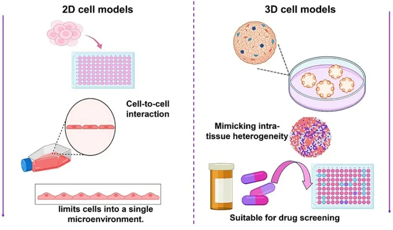
Historically, scientists have relied on two-dimensional (2D) cell culture models for drug toxicity testing because they are easy-to-use and highly scalable. However, there is increasing evidence that three-dimensional (3D) cell culture models may have improved predictivity due to better physiological relevance. Many researchers are now asking themselves, should I use 2D or 3D cell culture models for drug toxicity testing?
In this article, we'll compare 2D to 3D cell culture models for toxicity testing so you can choose the best approach for your preclinical studies.
Why In Vitro Models Matter in Drug Toxicity Testing
In vitro models are essential tools in drug toxicity screening because they enable rapid, cost-effective evaluation of compound safety before animal studies. These systems allow researchers to:
- Screen large numbers of compounds efficiently
- Identify cytotoxic or organ-specific toxic effects early
- Investigate mechanisms of toxicity at the cellular level
- Reduce reliance on animal testing
The usefulness of these assays, however, relies heavily on how predictive they are of human drug toxicity. Many factors influence this including how well your models resemble human biology. This is where the question of 2D vs 3D cell culture models for toxicity testing becomes critically important.
Commonly Used 2D and 3D Models for Drug Toxicity Testing
Selecting appropriate experimental models is essential for accurate drug toxicity assessment. Both 2D and 3D in vitro models are widely used to evaluate different types of toxicity, including hepatotoxicity, cardiotoxicity, nephrotoxicity, and neurotoxicity. Each model type offers distinct advantages depending on the application.
Common 2D Models in Drug Toxicity Testing
2D cell culture models remain the standard for early-stage screening due to their simplicity, scalability, and reproducibility.
Table 1. Widely Used 2D Models
| Type | Representative Cell Models | Key Features / Applications |
|---|---|---|
| Hepatotoxicity Models |
|
Widely used for liver toxicity screening, drug metabolism studies, and enzyme induction (CYP450); suitable for early DILI risk assessment |
| Cardiotoxicity Models |
|
Used for evaluating cytotoxicity, mitochondrial dysfunction, and electrophysiological effects (e.g., QT prolongation risk) |
| Nephrotoxicity Models |
|
Commonly used for assessing renal tubular toxicity, oxidative stress, and drug-induced kidney injury |
| Neurotoxicity Models |
|
Suitable for studying neurotoxicity, neuronal differentiation, and neurodegenerative mechanisms |
| Pulmonary Toxicity Models |
|
Used for inhalation toxicity, oxidative stress, and inflammatory response studies |
| Gastrointestinal Toxicity Models |
|
Commonly used for intestinal toxicity, permeability, and drug absorption studies |
| General Cytotoxicity Models |
|
High-throughput cytotoxicity screening and general cell viability assays |
Advantages of 2D Models in Toxicity Testing
Despite their drawbacks, 2D cell culture models are still widely used in academia and industry for toxicity testing. Here are some of the main reasons why:
- High throughput: 2D models are great for initial screening of large libraries of compounds.
- Easy to use: Many 2D cell assays are simple and can be easily reproduced.
- Cost-effective: 2D cell cultures are much cheaper to operate than their 3D counterparts.
- Mechanistic studies:2D models are ideal for toxicity studies that focus on cellular mechanisms.
Limitations of 2D Models for Drug Toxicity Assessment
While widely used, 2D models have significant limitations that can affect the accuracy of toxicity predictions.
- Lack of tissue architecture: Cells in 2D culture lack key features of normal tissue structure.
- Altered cell behavior: Gene expression patterns, differentiation states, and metabolic activity can be altered in 2D models.
- Limited cell-cell interactions: Reduced communication between cells affects biological responses.
- Poor clinical predictability:
These limitations can result in false positive or false negative toxicity outcomes, reducing translational relevance.s a result, relying solely on 2D models may not provide sufficient insight into human drug safety.
Common 3D Models in Drug Toxicity Testing
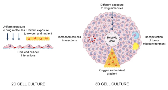
3D cell culture models are increasingly used to improve the predictive power of in vitro toxicity testing by better mimicking human tissue structure and function.
Table 2. Widely Used 3D Models
| Type | Representative 3D Models | Key Features / Applications |
|---|---|---|
| Hepatotoxicity Models |
|
Mimic liver architecture and metabolic activity; widely used for drug-induced liver injury (DILI) assessment and long-term toxicity studies |
| Cardiotoxicity Models |
|
Enable functional readouts such as contractility and electrophysiology; suitable for QT prolongation and cardiotoxicity prediction |
| Nephrotoxicity Models |
|
Recapitulate nephron structure and function; used for drug-induced kidney injury and transport studies |
| Neurotoxicity Models |
|
Reflect complex neural networks; used for neurodevelopmental toxicity and neurodegeneration studies |
| Pulmonary Toxicity Models | Lung organoids | Mimic airway structure and breathing dynamics; useful for inhalation toxicity and fibrosis studies |
| Tumor-related Toxicity Models |
|
Capture tumor heterogeneity and drug penetration; used for efficacy-toxicity balance evaluation |
| Multi-organ Models |
|
Advantages of 3D Models in Drug Toxicity Testing
Similar to how 2D cell cultures have been used for decades, 3D cell culture models are gaining traction for many applications in drug toxicity testing. Here are some advantages of 3D cell culture models for toxicity testing:
- They better represent how cells are organized in the human body.
- Cells interact with each other and the extracellular matrix in 3D models. This is important for eliciting proper biological responses to drugs.
- 3D cell cultures have been shown to have higher enzyme activity that is more predictive of liver toxicity.
- More predictive of organ-specific toxicity
- Drug response is more predictive of in vivo responses due to penetration gradients and drug diffusion.
These advantages make 3D cell models for drug toxicity assessment a powerful tool for improving predictive accuracy.
Limitations of 3D Models
Despite their benefits, 3D models also present certain challenges:
- Increased complexity: Establishing and maintaining 3D cultures requires specialized expertise.
- Higher cost: Materials, reagents, and equipment are typically more expensive.
- Lower throughput: Compared with 2D systems, scaling up experiments can be more difficult.
- Standardization challenges: Protocols may vary across laboratories, affecting reproducibility.
Therefore, while 3D models offer improved realism, they must be carefully implemented to ensure reliable results.
Key Differences Between 2D and 3D Cell Culture Models
The fundamental differences between 2D and 3D models directly influence their performance in toxicity testing.
| Feature | 2D Cell Culture Models | 3D Cell Culture Models |
|---|---|---|
| Cell structure | Flat monolayer | Three-dimensional architecture |
| Cell interactions | Limited | Physiologically relevant |
| Microenvironment | Artificial | Mimics in vivo conditions |
| Drug response | Often exaggerated | More realistic |
| Predictive power | Lower | Higher |
| Throughput | High | Moderate |
| Cost and complexity | Low | Higher |
This comparison highlights why 3D cell culture toxicity testing is gaining increasing attention as a more predictive alternative to traditional models.
When to Use 2D and 3D Models in Toxicity Testing
Choosing between 2D and 3D models depends on the specific goals of the study.
Choose 2D cell culture models when:
- You need to screen large numbers of compounds
- You are testing toxicity in the early stages of drug discovery
- You are interested in studying cellular mechanisms
- You are limited by budget/research time
Choose 3D cell culture models when:
- You are studying organ-specific toxicity
- You want to test longer-term or repeated dosing of drugs
- You want to confirm/validate toxicity findings from 2D assays
- You require more physiologically relevant models
In reality, you shouldn't have to pick between 2D vs 3D cell culture models. Most researchers will:
- Use 2D models for initial screening
- Use 3D cell cultures for validation and translation
- Use in vivo models and pharmacokinetics data to generate a more complete understanding of your compound.
By using a combination toxicity testing models, you'll have a better chance of making the right decision with respect to a drug's toxicity early in the drug discovery process.
A Final Word
2D and 3D cell culture models can both be useful for toxicity testing. 2D models are great for high-throughput screening and initial toxicity assessments, but they lack physiological relevance. 3D cell culture models, on the other hand, provide a more physiologically relevant system for predicting human drug toxicity.
It is important to understand the pros and cons of each model so you can make an informed decision about which one will work best for your studies. Instead of choosing one model over the other, you should use both 2D and 3D toxicity testing models in conjunction with each other.
Creative Bioarray Relevant Recommendations
| Products & Services | Description |
|---|---|
| Safety Evaluation Services | With years of expertise, Creative Bioarray serves as a trusted partner in safety evaluation, specializing in preclinical toxicology testing. Our comprehensive testing platform supports applications throughout all stages of product development. |
| In Vivo Toxicity Study | Creative Bioarray's GLP toxicology studies include administration routes ranging from acute to chronic. With internal resources, we can also provide a full range of toxicological assessments. |
References
- Dave R, Pandey K, et al. Leveraging 3D cell culture and AI technologies for next-generation drug discovery. Cell Biomaterials, 2025; 1
- Fontana F, Raimondi M, et al. Three-Dimensional Cell Cultures as an In Vitro Tool for Prostate Cancer Modeling and Drug Discovery. Int J Mol Sci. 2020 Sep 16;21(18):6806.