RKO
Cat.No.: CSC-C9242W
Species: Homo sapiens (Human)
Source: Intestine; Colon
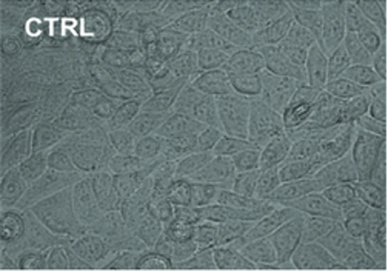
Morphology: epithelial
- Specification
- Background
- Scientific Data
- Q & A
- Customer Review
RKO is a line of human colorectal carcinoma cells. Established by Michael Brattain et al. from a primary colon carcinoma, RKO cells behave like most cancer cells morphologically (they are epithelial-like) and will grow aggressively in vitro as well as in xenograft assays. However, what sets RKO apart from other cancer cell lines is that it has well-defined mutations and epigenetic alterations. RKO is a microsatellite unstable (MSI-H) cell line due to epigenetic silencing of MLH1 via promoter hypermethylation. Therefore, RKO can be used as a model to study the "CIMP" pathway. Additionally, RKO has the BRAF (V600E) mutation and retains wild-type p53 expression, making it valuable for studying how oncogenic signaling interacts with wildtype tumor suppressor signaling. For this reason, it has become a popular cell line for performing high-throughput drug screens as well as DNA repair and gene editing studies because of its well-defined and understood behavior and sequenced genome.
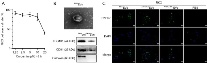
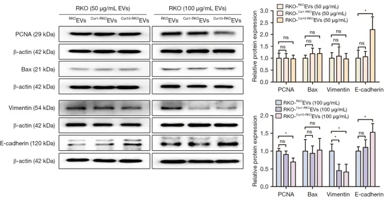
EVs Isolated from Cur-Medium Inhibited RKO Cell Proliferation
Curcumin (Cur) is a natural phytochemical that is expected to become an indispensable drug for the treatment of colorectal cancer. A comprehensive understanding of the anti-tumor mechanism of Cur will provide a better reference for its clinical application. Xu's team aimed to examine the effects of extracellular vesicles (EVs) isolated from Cur-medium on RKO colorectal cancer cell proliferation, apoptosis, and migration.
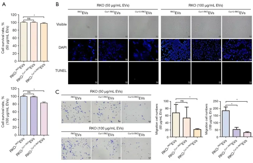
RKO cells treated with 1.25-20 µM curcumin (Cur) for 48 hours showed survival rates of 91.23%±4.13% to 39.64%±3.26% (Fig. 1A), indicating dose-dependent viability reduction. They selected 1.25 µM and 10 µM Cur for further study. EVs were isolated from RKO cells treated with 1.25 µM and 10 µM Cur (Cur1-RKOEVs and Cur10-RKOEVs) (Fig. 1B). PKH67-labeled EVs were taken up by RKO cells (Fig. 1C). At 50 µg/mL, Cur1-RKOEVs and Cur10-RKOEVs had minimal impact on RKO cell survival (100.13%±1.48% and 97.07%±1.28%) or PCNA expression (Fig. 2, 3). However, at 100 µg/mL, Cur10-RKOEVs significantly reduced survival to 81.76%±1.84% and inhibited PCNA expression, while Cur1-RKOEVs had no significant effect (98.85%±1.51%) (Fig. 2, 3). This suggests that high-dose Cur-derived EVs inhibit RKO cell proliferation.
Ask a Question
Write your own review
- You May Also Need

Description: Established from an adenocarcinoma located in the ascending colon of a 68 year-old male patient. The adenocarcinoma was well differentiated and determined to be Dukes' stage B. Imperial College ...

Description: This is one cell line out of a series of colon carcinoma cell lines established by PD Dr. Michael Linnebacher.
Description: Rectal carcinoma from a Japanese patient. Cell growth is slow.
Description: The C80 cell line was established from a 69-year old male patient with a moderately well differentiated adenocarcinoma of the rectum classified as Dukes' stage D.

Description: Species: human - male, 70 years old, CaucasianTumorigenecity: yes, in nude miceIsoenzyme: G6PD,B;PGM1,1-2;PGM3,1-2;6PGD,A;ES-D,1-2;PEP-D,1Histopathology: adenocarcinomaNote: CSAp negative (CSAp-); ...
- Adipose Tissue-Derived Stem Cells
- Human Neurons
- Mouse Probe
- Whole Chromosome Painting Probes
- Hepatic Cells
- Renal Cells
- In Vitro ADME Kits
- Tissue Microarray
- Tissue Blocks
- Tissue Sections
- FFPE Cell Pellet
- Probe
- Centromere Probes
- Telomere Probes
- Satellite Enumeration Probes
- Subtelomere Specific Probes
- Bacterial Probes
- ISH/FISH Probes
- Exosome Isolation Kit
- Human Adult Stem Cells
- Mouse Stem Cells
- iPSCs
- Mouse Embryonic Stem Cells
- iPSC Differentiation Kits
- Mesenchymal Stem Cells
- Immortalized Human Cells
- Immortalized Murine Cells
- Cell Immortalization Kit
- Adipose Cells
- Cardiac Cells
- Dermal Cells
- Epidermal Cells
- Peripheral Blood Mononuclear Cells
- Umbilical Cord Cells
- Monkey Primary Cells
- Mouse Primary Cells
- Breast Tumor Cells
- Colorectal Tumor Cells
- Esophageal Tumor Cells
- Lung Tumor Cells
- Leukemia/Lymphoma/Myeloma Cells
- Ovarian Tumor Cells
- Pancreatic Tumor Cells
- Mouse Tumor Cells