Common Immunohistochemistry Stains and Their Role in Cancer Diagnosis
Did you know? Over 90% of cancer diagnoses rely on immunohistochemistry (IHC) to pinpoint tumor origin and guide treatment. But how do these tiny stains unlock such critical answers?
Core Principles of IHC
The main principle of IHC is the specific binding between antibodies and antigens. Antibodies bind to specific antigens in tissue samples and are visualized through chromogenic or fluorescent labeling, allowing for the detection and localization of specific antigens in tissues. The procedure usually involves antigen retrieval, antibody incubation, and chromogenic/fluorescent detection.
IHC has been widely applied in medical diagnostics, pathological research, and basic science. It has become an irreplaceable technique in the diagnosis of cancers. It is not only used to differentiate different types of tumors (such as adenocarcinoma, squamous cell carcinoma, and sarcoma) but also to determine the aggressiveness, metastatic potential, and treatment response of tumors.
Applications of IHC in Cancer
Cancer diagnosis
Specific protein expression in tumor tissues can be detected by IHC staining, for example, Ki-67 (proliferation marker), EGFR (epidermal growth factor receptor), etc. These markers can be used for diagnosis, as well as for prognosis evaluation and treatment selection.
Tumor microenvironment research
Histological and IHC staining can be used to analyze the types of cells in the tumor microenvironment, intercellular interactions, and changes in signaling pathways.
Treatment response evaluation
Histological staining can be used to detect morphological changes, apoptosis, and necrosis in tumor tissues to evaluate the efficacy of cancer treatments, such as chemotherapy, radiotherapy, and other therapeutic methods.
Nanomedicine applications
IHC can be applied in nanomedicine to study the targeting efficiency of nanoparticles, their therapeutic effects, and their toxicity evaluation.
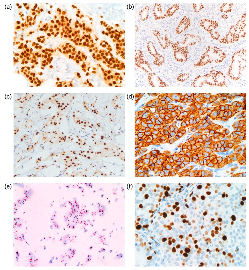
Common IHC Markers in Cancer Diagnosis
In IHC-based cancer diagnostics, first-line markers serve as essential tools for initial tumor classification. These markers exhibit high sensitivity and broad expression profiles, aiding pathologists in rapidly identifying tumor origin and histological type.
1. Epithelial tumor markers
Cytokeratins (CKs), particularly CK7 and CK20 combinations, help differentiate carcinomas (e.g., lung adenocarcinoma vs. colorectal cancer). CKs remain detectable in necrotic tissue but may cross-express in non-epithelial tumors (e.g., melanoma, lymphoma).
- CK7+/CK20-: Common in lung adenocarcinoma, breast cancer, ovarian cancer.
- CK7-/CK20+: Suggests colorectal or gastric adenocarcinoma.
- CK7+/CK20+: May indicate urothelial or pancreatic cancer.
- CK7-/CK20-: Possible hepatocellular carcinoma or certain neuroendocrine tumors.
- CK5/6: Expressed in squamous cell carcinoma (e.g., lung) and basal-like breast cancer.
Special Staining Patterns:
- Renal oncocytoma: CK20 shows perinuclear dot-like staining.
- Seminoma: CK exhibits discontinuous membranous or globular cytoplasmic staining.
2. Neuroendocrine tumor markers
Neuroendocrine neoplasms (NENs) secrete hormones and arise in various sites (e.g., lung, pancreas, GI tract). Diagnosis requires combined morphology and IHC.
- Chromogranin A (CgA): High specificity but may be weak/absent in poorly differentiated tumors.
- Synaptophysin (Syn): Highly sensitive, widely used for NENs, especially metastatic cases.
- CD56: Sensitive but nonspecific; requires corroboration.
- INSM1: Novel marker with high specificity for poorly differentiated neuroendocrine carcinomas.
Note:
- ~20% of non-NEN lung tumors may express neuroendocrine markers (mimic NENs).
- Merkel cell carcinoma is the only CK7−/CK20+ neuroendocrine tumor.
3. Melanoma markers
Melanoma diagnosis requires multiple markers due to limited sensitivity/specificity of single markers.
Screening Markers:
- S100: >95% sensitivity but low specificity (also stains schwannomas, sarcomas).
- SOX10: Nuclear staining; more sensitive for desmoplastic melanoma.
Confirmatory Markers:
- Melan-A/MART-1: ~80% sensitivity; may be false-positive in adrenal/renal tumors.
- HMB45: High specificity but negative in desmoplastic melanoma.
- PRAME: Novel marker for distinguishing benign vs. malignant melanocytic lesions.
- Pitfall: Metastatic melanoma may lose markers; use CK/CD45 to exclude other tumors.
4. Hematolymphoid markers
Lymphoma diagnosis relies on multiple markers to differentiate B-cell, T-cell, and Hodgkin lymphomas.
Pan-Lymphocyte Markers:
- CD45 (LCA): Marks most lymphocytes but negative in Hodgkin-Reed-Sternberg (HRS) cells.
B-cell Markers:
- CD20: Key for B-cell lymphomas (rituximab target); post-treatment clones may lose CD20.
- PAX5: Nuclear expression; may cross-react with some T-cell lymphomas/carcinomas.
T-cell Markers:
- CD3: Pan-T-cell marker; subset analysis via CD4/CD8.
Plasma Cell Marker:
- CD138: Marks plasma cells but may express in carcinomas.
5. Mesenchymal tumor markers
Soft tissue sarcomas require markers to determine muscle, vascular, or neural origin.
- Vimentin: Broad mesenchymal marker; low specificity.
- Desmin/SMA: Marks smooth muscle/rhabdomyosarcoma.
- CD31/CD34: Vascular tumor markers.
Second-line markers are highly specific markers used to further pinpoint the origin of tumors based on initial findings from first-line markers. These markers are typically transcription factors or tissue-specific proteins and often exhibit nuclear staining patterns. They are employed to resolve challenging cases or identify the primary site of metastatic cancers. Common second-line markers include:
- TTF-1: Thyroid/lung adenocarcinoma (80–90% lung ADC+); may express in neuroendocrine carcinomas.
- GATA3: Sensitive for breast/urothelial cancer (high in ER+ breast cancer); may stain lymphomas/mesotheliomas.
- CDX2: GI adenocarcinoma marker (>90% colorectal cancer+); expression correlates with differentiation.
- PAX8: Thyroid/kidney/ovarian/endometrial cancer marker; may cross-react with PAX6.
This comprehensive approach ensures precise cancer classification and personalized treatment strategies.
Creative Bioarray Relevant Recommendations
| Products & Services | Description |
|---|---|
| Immunohistochemistry (IHC), Immunofluorescence (IF) Service | Creative Bioarray offers a comprehensive IHC service from project design, marker selection to image completion and data analysis. |
| Histology Services | Creative Bioarray offers tissue processing, embedding, sectioning, and staining, along with set of histological examination services. |
References
- Sanguedolce F, Zanelli M. Immunohistochemistry in the Diagnosis of Primary and Secondary Cancers. In: Rezaei, N. (eds) Handbook of Cancer and Immunology. Springer, Cham. 2022
- Hacking SM, Yakirevich E, et al. From Immunohistochemistry to New Digital Ecosystems: A State-of-the-Art Biomarker Review for Precision Breast Cancer Medicine. Cancers. 2022. 14(14):3469.