Karyotyping (G-Banded) Service
- Service Details
- Features
- Application
- FAQ
- Explore Other Options
Chromosomal instability remains one of the leading causes of failed cell-based research, regulatory delays, and inconsistent experimental outcomes. G-banded karyotyping is the gold standard method for detecting numerical and structural chromosomal abnormalities above 5 Mb.
Creative Bioarray provides high-resolution, species-specific karyotyping services to ensure genomic stability and compliance in research and therapeutic applications.
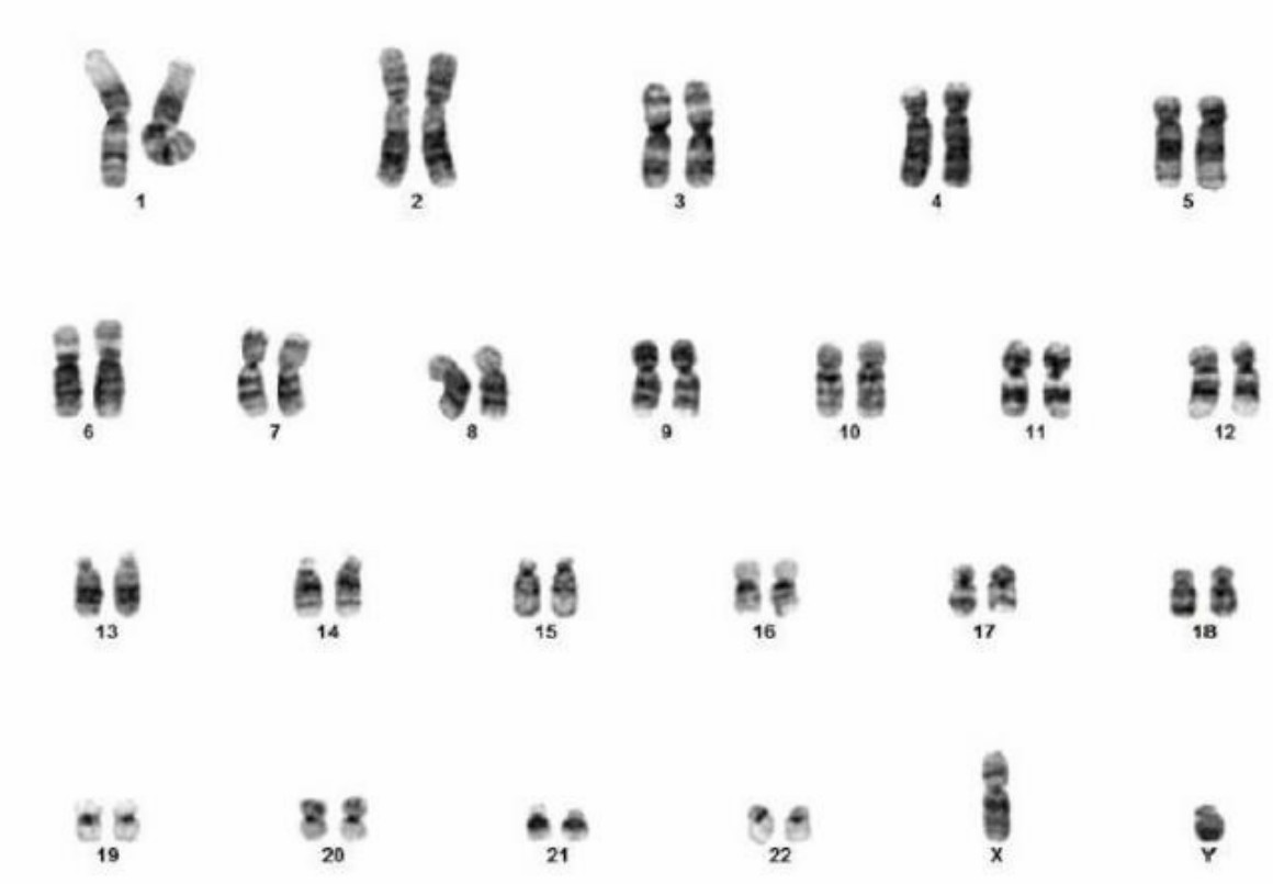
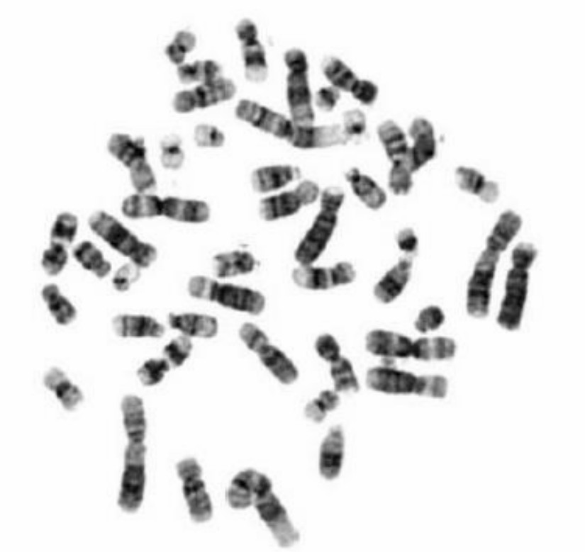
Fig. 1. The metaphase chromosome spread (right) and corresponding karyotype (left) of a normal human male.
Use karyotype (G-Banded) analysis to detect:
Microscopic genomic abnormalities> 5Mb
- Aneuploidy
- Inversions
- Duplications/deletions
- Translocations
- >10% Mosaicism
Our Karyotyping (G-Banded) Service
Workflow
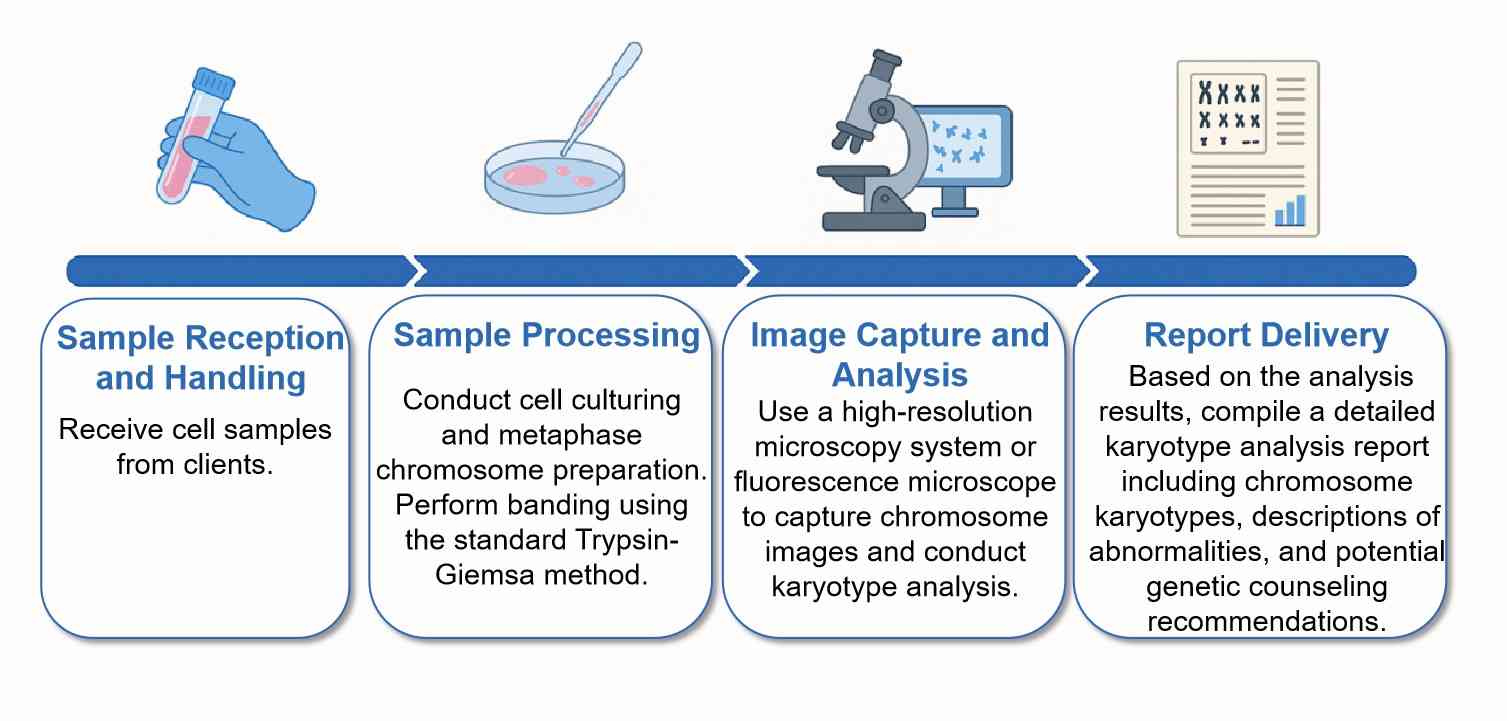 Fig. 1. The metaphase chromosome spread (right) and corresponding karyotype (left) of a normal human male.
Service
Species-Specific Karyotyping
- Human Karyotyping Service
- Mouse Karyotyping Service
- Rat Karyotyping Service
- Rare Species Karyotyping Service (e.g., Sheep, Cow, Pig, Dog, Hamster, Fish)
Cancer Cells Karyotyping
- Complex analysis for cancer cell lines and cases with unstable karyotypes
Cell Line Karyotyping
- Assessment of genomic stability and chromosomal changes
- Detection of inversions, translocations, and aneuploidy
- Culture development is available for NK Cells, iPSCs, T-Cells, and whole blood
Reporting Standards
- Analysis of 20 metaphase spreads minimum
- 30–100+ cell counting upon request
- Representative high-resolution karyotype images
- Raw metaphase image files included
- Optional FISH confirmation available
Why Choose Our Karyotyping (G-Banded) Service?

Diverse Species Expertise
We analyze multiple species, customizing methods for varied biological needs.

Advanced Techniques
High-detail G-banding and multicolor FISH probes set industry standards.

Comprehensive Reporting
Clear and detailed reports provide full understanding of karyotyping results.

Tailored Service
Personalized solutions with continuous support to meet specific needs.
When Should You Use Our Karyotyping (G-Banded) Service?
You may need karyotyping analysis if you are facing any of the following situations:
- Unexpected changes in cell behavior
Abnormal growth rates, altered morphology, or unstable differentiation in cultured cells.
- Long-term cell culture or high passage numbers
To verify genomic stability before publication, project continuation, or downstream applications.
- Stem cell or iPSC expansion
To monitor chromosomal integrity during reprogramming and prolonged culture.
- Cancer cell line characterization
To identify complex structural rearrangements and chromosomal instability.
- Cell bank validation or quality control
To confirm chromosomal normality before cryopreservation or distribution.
- Breeding or species cytogenetic studies
To compare chromosome patterns and detect structural variations.
FAQ
1. How long does the karyotyping process take?
The entire process, from sample receipt to report delivery, usually takes7-10 Business Days, depending on the sample type and complexity of the analysis.
2. What samples can be used for Karyotyping (G-Banded) testing?
We accept peripheral blood, amniotic fluid, chorionic villus tissue, bone marrow, and other tissue samples. The specific choice depends on the testing purpose and client requirements.
3. What are the sample requirements?
Generally, 2-3 milliliters of peripheral blood are needed (collected in heparin anticoagulant tubes), more than 15 milliliters of amniotic fluid (in a sterile container), and more than 20 milligrams of chorionic villus tissue (preserved in transport medium). For other tissue samples, please consult professionals. Live cell samples should be submitted for testing as soon as possible with cold transport, while fixed cell samples should be handled according to laboratory guidelines.
4. What does the report include?
The report includes a chromosome karyotype image, detailed descriptions of chromosomal abnormalities, karyotype formula, and genetic diagnostic opinions. Additionally, raw microscope image files are provided for client reference.
Explore Other Options