
KG-1
Cat.No.: CSC-C8220L
Species: Homo sapiens (Human)
Source: Bone Marrow
Morphology: myeloblast-like
Culture Properties: Suspension
- Specification
- Background
- Scientific Data
- Q & A
- Customer Review
- Documents
CSF1PO: 7
D13S317: 11,12
D16S539: 10,11
D5S818: 13
D7S820: 8,10
THO1: 7,8
TPOX: 7,9
vWA: 14,19
The KG-1 cell line was derived from the bone marrow aspirate of a 59-year-old male patient who had initially presented with erythroleukemia that then progressed to acute myelogenous leukemia (AML). Microscopically, KG-1 cells exhibit considerable pleomorphism, with a predominance of myeloblasts and promyelocytes, which is consistent with the AML phenotype.
The availability of the KG-1 cell line has been highly valuable for researchers studying the molecular and cellular mechanisms underlying AML pathogenesis, as well as for evaluating potential therapeutic interventions for this aggressive form of leukemia. The cell line's properties, such as colony formation and responsiveness to growth factors, make it a useful in vitro model for investigating the dysregulation of myeloid differentiation and proliferation in AML.
Regulation of the Gene for CD34 in KG-1 Cells
CD34 is one of the best-characterized human hematopoietic stem cell antigens defined to date. CD34 expression is lost during hematopoietic development and is not found on mature peripheral blood cells. The control of CD34 expression was studied in the myeloblast cell line KG-1 as a model for the regulation of stem cell genes.
To study the effect of differentiation on the steady state level of CD34 mRNA in KG-1 cells, KG-1 cells were induced towards macrophages by treatment with 10-7 mol/L TPA and 1.6 μg/mL ionomycin at various times. Total RNA was isolated from uninduced and induced KG-1 cells and probed with the 1.5 kb CD34 cDNA. CD34 mRNA was expressed at high levels in uninduced KG-1 cells and was reduced 10-fold after 24 hours of induction. Very little CD34 mRNA remained at 48 hours (Fig. 1). To ascertain whether the observed down-regulation of CD34 is due to transcriptional or post-transcriptional mechanisms, nuclear run-on transcription assays were done using nuclei isolated from uninduced KG-1 cells and KG-1 cells induced with 10-8 mol/L TPA and 0.16 μg/mL ionomycin for 24 hours. In two independent experiments, the rate of CD34 transcription did not change from its uninduced level (Fig. 2). In contrast, transcription of the c-myc gene consistently decreased twofold upon induction (Fig. 2). The rate of transcription of actin reproducibly increased 3.5-fold in induced KG-1 cells (Fig. 2). CD18 transcription also increased, to a variable degree, upon induction (Fig. 2).
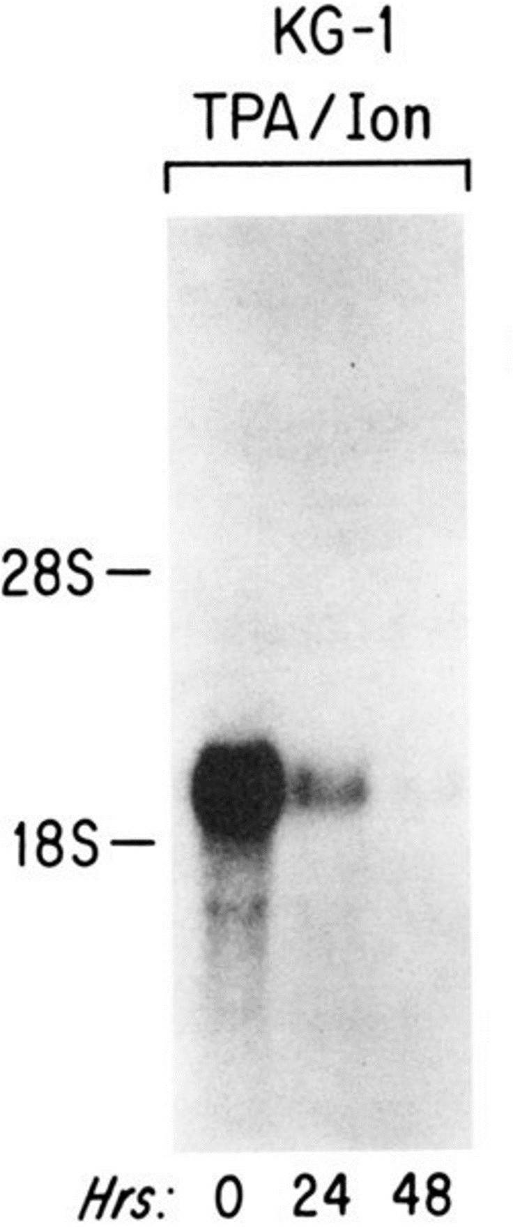 Fig. 1 Northern blot analysis of the effect of TPA and ionomycin on CD34 mRNA level in KG-1 cells. (Satterthwaite AB, et al., 1990)
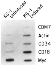 Fig. 2 Nuclear run-on analysis of the effect of TPA and ionomycin on transcription rates in KG-1 cells. (Satterthwaite AB, et al., 1990)
Effects of Different Autophagy Inhibitors on KG-1 Leukemia Cells
A small number of current autophagy inhibitors may have beneficial effects on AML patients. However, there is a strong need to figure out which settings should be activated or inhibited in the autophagy pathway to prevail against drug resistance and also to improve current treatment options in leukemia. Compare the effects of well-known inhibitors of autophagy (as 3-MA, BafA1, and HCQ) in leukemia KG-1 cells exposed to arsenic trioxide (ATO) and/or all-trans retinoic acid (ATRA).
The 50% inhibitory concentrations (IC50) of ATO and IC30 of ATRA were determined using an MTT assay (Fig. 3). The IC50 for ATO was 1.61 μM in KG-1 cells. IC30 for ATRA was approximately 700 nM in KG-1 cells. The combination of ATO with ATRA further reduced the cell proliferation (up to 60%) in KG-1 and HL-60 cell lines, in comparison with each monotherapy (Fig. 3). Therefore, ATO/ATRA is known as a more potent inhibitor of the viability of leukemia cells compared to each monotherapy.
The occurrence of autophagy was further evaluated by the mRNA expression of typical ATGs using real-time PCR. The up-regulated mRNA expressions of BECN1, LC3B, P62, ATG12, and ULK in KG-1 ATO/ATRA treated cells compared to the control cells (Fig. 4). After the treatment of KG-1 cells for 48 h, ATO/ATRA in combination with HCQ, up-regulated the mRNA expression of LC3B, P62, CASP3, ATG12, and ULK; whereas they down-regulated BECN1 and ATG7 compared to the controls. After the treatment of HL-60 cells for 48 h, ATO/ATRA combined with HCQ up-regulated the mRNA expressions of LC3B, P62, CASP3, ATG12, and ATG7; whereas they down-regulated BECN1 and ULK compared to the controls.
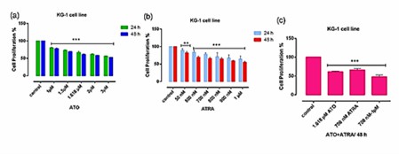 Fig. 3 Cell proliferation in KG-1 cells. (Haghi A, et al., 2021)
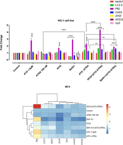 Fig. 4 The mRNA expression in KG-1 cell lines. (Haghi A, et al., 2021)
Ask a Question
Write your own review
- You May Also Need

Description: Established in 2007 from the bone marrow mononuclear cells of an 82-year-old Japanese man with diffuse large B-cell lymphoma in the leukemic phase

Description: Established from the bone marrow of a 28-year-old man who developed the terminal leukemic phase of lymphosarcoma in 1976

Description: Established from the pleural effusion of a 24-year-old woman with recurrent anaplastic large cell lymphoma (ALCL); cells were described to clonally derive from T-lineage lymphoid cells and to be ...

Description: Established from a 37-year-old man at second (refractory/terminal) relapse of Hodgkin lymphoma (nodular sclerosing -> lymphocyte depleted/stage IIISA -> stage IV) after both combined chemo- and ...

Description: Established from the peripheral blood of a 10-year-old Caucasian boy with acute lymphoblastic leukemia (pre B-ALL) at diagnosis in 1993

Description: Established from the peripheral blood of a 69-year-old woman with B-CLL (chronic lymphocytic leukemia) prior treatment by EBV-transformation in 1988; cell line may represent rather a B-lymphoblastoid ...
- Adipose Tissue-Derived Stem Cells
- Human Neurons
- Mouse Probe
- Whole Chromosome Painting Probes
- Hepatic Cells
- Renal Cells
- In Vitro ADME Kits
- Tissue Microarray
- Tissue Blocks
- Tissue Sections
- FFPE Cell Pellet
- Probe
- Centromere Probes
- Telomere Probes
- Satellite Enumeration Probes
- Subtelomere Specific Probes
- Bacterial Probes
- ISH/FISH Probes
- Exosome Isolation Kit
- Human Adult Stem Cells
- Mouse Stem Cells
- iPSCs
- Mouse Embryonic Stem Cells
- iPSC Differentiation Kits
- Mesenchymal Stem Cells
- Immortalized Human Cells
- Immortalized Murine Cells
- Cell Immortalization Kit
- Adipose Cells
- Cardiac Cells
- Dermal Cells
- Epidermal Cells
- Peripheral Blood Mononuclear Cells
- Umbilical Cord Cells
- Monkey Primary Cells
- Mouse Primary Cells
- Breast Tumor Cells
- Colorectal Tumor Cells
- Esophageal Tumor Cells
- Lung Tumor Cells
- Leukemia/Lymphoma/Myeloma Cells
- Ovarian Tumor Cells
- Pancreatic Tumor Cells
- Mouse Tumor Cells