Cell Surface Marker Validation Service
Cell surface proteins have a wide range of biological functions, including mediation of cell-cell communication and responses to external signals such as the presence of pathogens or chemical messangers. And they are often used as lineage-specific markers, which define phenotypic and functional differences between cell types, and between normal and diseased cells, such as cancer cells. Rapid characterization of the cell surface markers could not only lead to identification and development of new diagnostic markers and theapeutic targets, but also provide insight into the basic biology of diesease. The ability to obtain broad cell surface protein profiles would thus be of great value in a wide range of fields.
Creative Bioarray provides state-of-the-art cell surface marker validation service to support all researchers, biotech and pharmaceutical companies in the progress of drug development. We provide the full cell surface markers validation services from experimental design to specimen progressing, acquisition, and data analysis.
Stem Cell
Over three decades of stem cell research are currently culminating in remarkable clinical results, highlighting not only the broad applicability of these approaches, but also the immense future prospects of cell and gene therapy using adult stem cells and somatic derivative of pluriopotence stem cells (PSCs). The combined disciplines of cell and gene therapy and tissue engineering, broadly known as regenerative medicine, have the potential to revolutionize the treatment of diseases and injuries.
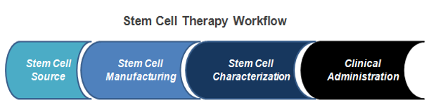
So in the rapidly growing field of stem cell therapy, there is a need for characterization of stem cells. For example, mesenchymal and tissue stem cell committee of the International Society for Cellular Therapy (ISCT) has proposed minimal criteria to define human MSCs. MSCs must be plastic adherent, express CD105, CD73 and CD90 and lack expression of CD45, CD34, and CD14 Or CD19 and HLA-DR surface molecules, as well as be capable of differentiation to osteogeneic, adipogenic and chondrogenic lineages.
Table 1. Summary of Minimal Criteria* to Identify MSC
| Phenotype | Positive (≥95%+) | Negative (≤2%+) |
| CD105 | CD45 | |
| CD73 | CD34 | |
| CD90 | CD14 | |
| CD79 | ||
| HLA-DR |
* Proposed by International Society for Cellular Therapy (ISCT)
Immune Cells and Primary Cells
Immunopheotyping is a useful technique to analyze the immune cell subsets by detecting proteins expressed on the surface or wIthin the cell (intracellular) using antigen specific antibodies. In order to classify the cell types of interest, detecting multiple antigenes on the cells is often necessary. Primary cell surface maker validation is also very important, the makers have been linked to transmembrane transport and inter-cellular communication.
Table 2. Brief Summary of Immune Cell and Primary Cell Surface Markers
| Cell Type | Cell Surface Markers |
| B cell | CD1C, CD28, CD79, CD80, CD86, CR1, FCGR2A, ITGA3, MS4A1, NT5E |
| T cell | CD4, CD8, CD3, CD6, CD40LG, CTLA4, FAS, TNFRSF4 |
| Natural killer (NK) cell | CD2, CD7, CD96, CD244, CD247, IL12RB1, KLRs, NCAM1 |
| Macrophage/monocyte | CD33, CD63, CD69, CD70, CD74, DPP4, IL1R2, ITGA1, TNFRSF8 |
| Platelet | ITGA2B, ITGB3M SELP |
| Dendritic cell | CD1A, CD40, CD83, CD86, CD209, IL3RA, ITGAX |
| Granulocyte | C5AR1, CEACAM8, FCER1A, FCER2, FCGR3A |
| Endothelial cell | ENG, ICAM2, MCAM, NOS3, PECAM2, TEK, VCAM1, VWF |
| Epithelial cell | CD1D, EPCAM, KRT8, KRT18 |
| Smooth muscle cell | MYH9, MYH19, MYOCD |
| Fibroblast | ALCAM, COL1A1, COL1A2 |
Creative Bioarray provides Cell Surface Markers Validation Service, including but not limited to:
- Growth factor, cytokine, and chemokine receptors
- Cell-cell interaction molecules
- Antigen presentation and processing
- Stem cell markers
- T cell, B cell NK cell activation
- Analyze cell lines, primary cells, blood samples
- Discover new biomarkers
- Custom Panels are also available
Quotation and ordering
Our customer service representatives are available 24hr a day! We thank you for choosing Creative Bioarray at your preferred Cell Surface Markers Validation Service.
References
- Secunda, R., et al. "Isolation, expansion and characterisation of mesenchymal stem cells from human bone marrow, adipose tissue, umbilical cord blood and matrix: a comparative study." Cytotechnology 67.5 (2015): 793-807.
- Gedye, Craig A., et al. "Cell surface profiling using high-throughput flow cytometry: a platform for biomarker discovery and analysis of cellular heterogeneity." PloS one 9.8 (2014): e105602.
Explore Other Options