Immunohistochemistry (IHC), Immunofluorescence (IF) Service
- Service Details
- Workflow
- Features
- FAQ
- Explore Other Options
Immunohistochemistry (IHC) and Immunofluorescence (IF) are indispensable techniques in modern biomedical research. IHC employs enzyme-chromogen reactions on paraffin-embedded or frozen tissue sections to achieve precise antigen localization, establishing this technique as the gold standard for protein expression analysis in both diagnostic pathology and experimental research. In contrast, IF employs fluorophore-conjugated antibodies to enable fluorescent tagging of target proteins. This facilitates direct observation of protein spatial distribution at cellular and tissue levels. Both techniques are widely applied in histopathology, oncology, neuroscience, and related fields.
Should You Choose IHC or IF?
- IHC: Ideal for experiments requiring long-term observation and preservation, especially when sample stability is paramount. This method produces color-stable tissue sections, making them easy to preserve and analyze over extended periods.
- IF: Suited for experiments necessitating dynamic observation. With its high sensitivity and multicolor labeling capabilities, IF enables simultaneous observation of multiple target antigens, making it an ideal choice for studying complex intracellular protein interactions.
Our expert team provides tailored guidance and advice based on the specific requirements and objectives of your experiment.
Creative Bioarray's Comprehensive IHC and IF Services
IHC Services:
Chromogenic IHC services are suitable for routine pathological diagnosis and basic research involving protein localization analysis. Our professional team optimizes staining conditions according to your needs, ensuring accuracy and consistency of the staining results.
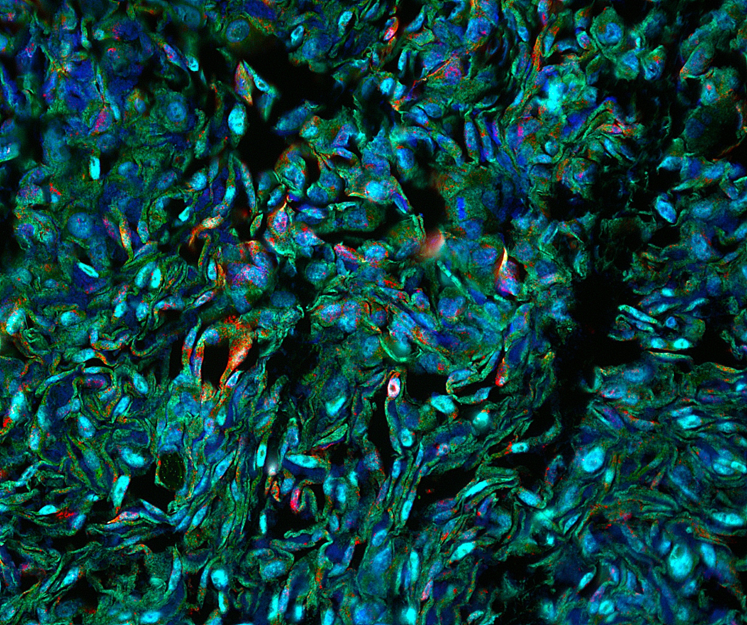
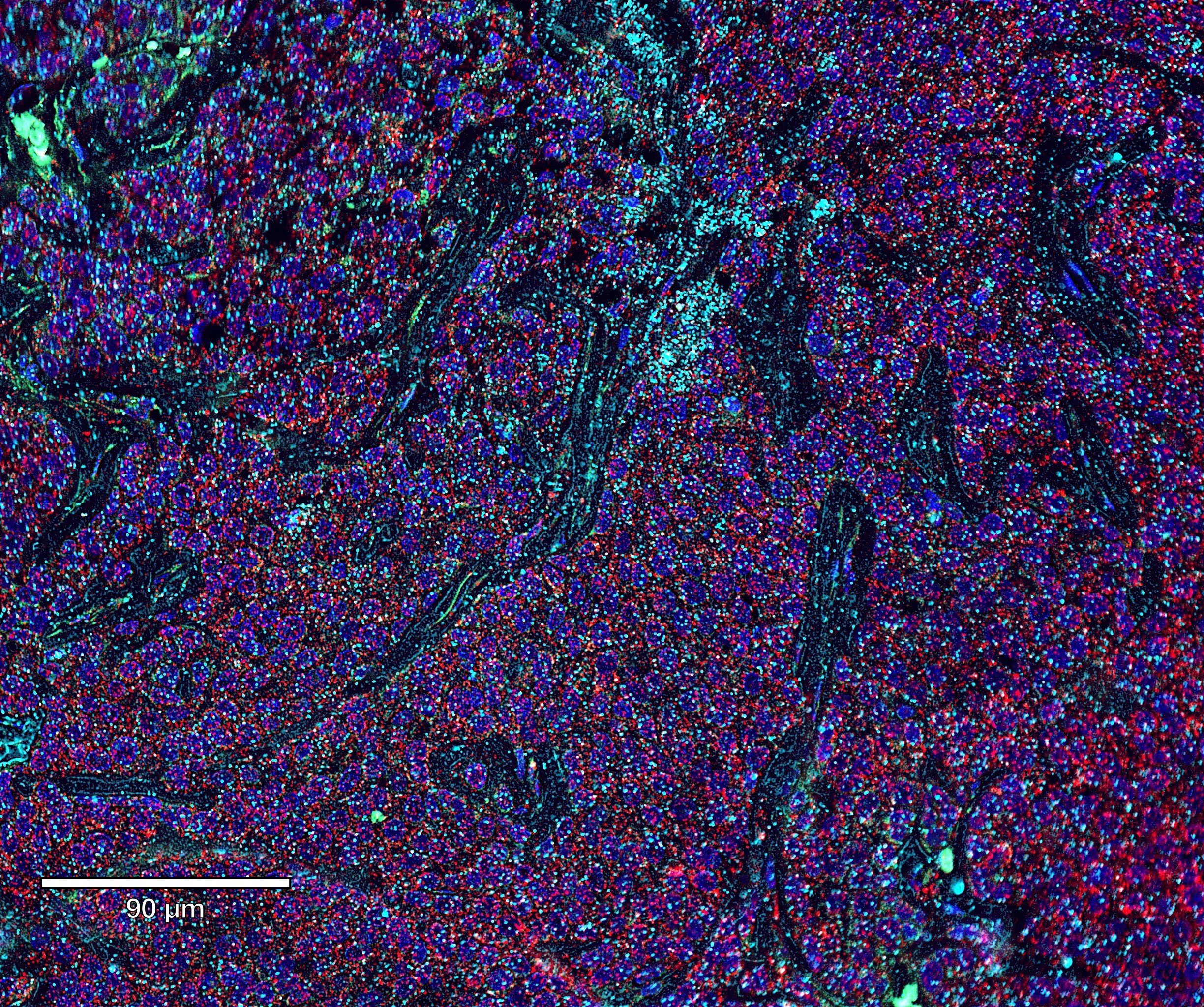
IF Services:
We offer a variety of fluorescent dye options, enabling the simultaneous labeling of multiple proteins to meet your research needs concerning complex molecular interactions and cellular structures. Additionally, our skilled technicians possess rich experience in fluorescence microscopy, providing you with high-quality image acquisition and analysis services.
- High-Quality Antibodies: We offer rigorously selected and validated antibodies.
- Platform Choice: Equipped with advanced IHC and IF detection platforms (Histopathology Platform).
- Control Tissues: We select appropriate positive and negative controls sourced from rigorously diagnosed samples.
- Experiment Condition Setup: We optimize parameters such as antibody concentration, incubation time, and temperature for different samples and antibodies.
- Comprehensive Reports: Provide detailed image and analysis reports to support further research decisions.
Capabilities of Our IHC and IF Service Platform:
- Analyze Multiple Biomarkers: Comprehensive analysis of multiple targets within the same tissue.
- Professional ROI Analysis Services: Precision analysis of specific regions within tissue sections based on research needs.
- Custom Multiplex Immunofluorescence Panels: Tailored multiplex immunofluorescence panels according to your research requirements.
- Image Analysis Technology: In-depth analysis of biomarker expression, cell phenotyping, and spatial distribution.
- Extensive Validation Projects: Ensures reliability and reproducibility of experimental results.
- Color and Fluorescent Single/Dual/Multiplex Detection: Accommodates complex experimental design needs.
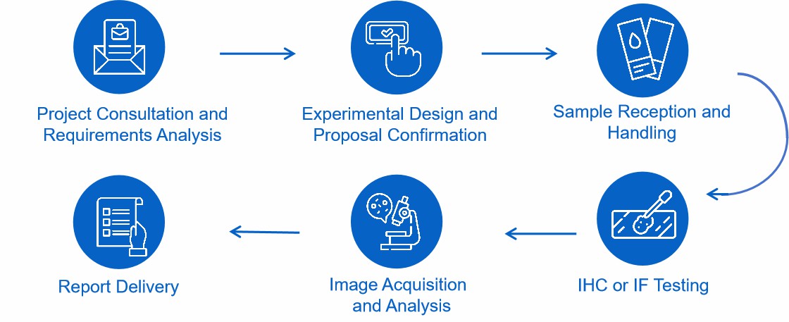
Workflow
Features

Professional Team
Our team comprises highly skilled technicians with extensive expertise and practical experience in the field of IHC and IF.

Advanced Equipment
Equipped with advanced automated stainers, fluorescence microscopes, and image analysis systems.

Customized Service Solutions
Provides flexible service options tailored to customer needs, including experimental design, antibody selection, and condition optimization.

Rapid Response
Upon contact, we promptly arrange for our expert technicians to communicate with you and respond swiftly to your needs.
FAQ
1. What are the sample requirements for IHC and IF testing?
IHC and IF samples typically include fresh or fixed tissue sections. To avoid antigen degradation and autolysis, fresh tissue samples must undergo immediate fixation after collection. Fixed tissue sections should be well-preserved and evenly sliced (generally 4-5 microns). Please mark the source, collection time and handling method of the sample to facilitate experiment and result analysis.
2. Can you provide raw data and image files from experiments?
Yes, we can provide original data and image files alongside your report to facilitate further analysis and study.
3. Do you offer antibody validation services?
Yes, we offer antibody validation services. All antibodies we use are rigorously screened and validated to ensure specificity and affinity.
4. How do you ensure the accuracy and reliability of experimental results?
We implement multiple measures to guarantee quality:
- Antibody Selection: We use only highly specific, high-affinity antibodies and have them tested their titer regularly.
- Standardized Processes: We have detailed SOPs on all the steps from sample handling and staining to microscopic observation to guarantee standardized processing.
- Control Setup: We use positive control (known target expressing tissues) and negative control (irrelevant antibodies or without primary antibodies) to guarantee system efficacy.
- Repeat Experiments: We do repeat the critical experiments for reproducibility.
- Data Analysis: We use professional image analysis software and professional technicians to interpret the data to minimize human error.
Explore Other Options