MT-2
Cat.No.: CSC-C6756J
Species: Homo sapiens (Human)
Source: Blood; Umbilical Cord Blood
Morphology: Lymphoid
- Specification
- Background
- Scientific Data
- Q & A
- Customer Review
The MT-2 cell line was derived from normal human cord leukocytes of a healthy donor by co-cultivation with leukemic cells from an adult T-cell leukemia (ATL) patient. This unique cell line offers a valuable model for studying certain aspects of T-cell biology and their therapeutic potential.
One significant characteristic of the MT-2 cell line is its ability to produce TGF-β, an immunosuppressive cytokine, which may contribute to the cell line's suppressive activity. Additionally, the MT-2 cell line has been shown to have the phenotypic and functional characteristics of human regulatory T cells (Tregs), suggesting that it may be used as a human Treg-like cell line for studies related to these cells. In addition to its potential role in Treg studies, the MT-2 cell line is also used to detect syncytium-inducing (SI) variants of HIV. In the MT-2 cell culture assay, MT-2 cells are cultivated with cell-free supernatants from HIV-infected PBMC cultures. The inoculated MT-2 cell cultures are then monitored for the development of a typical cytopathic effect, providing valuable insights into the behavior of HIV.
Melphalan Improves Toxicity of Arsenic Trioxide in Adult T-cell Leukemia/Lymphoma Cells
Adult T-cell leukemia/lymphoma (ATLL) is a hematologic neoplasm with poor prognosis. Melphalan is an alkylating anti-cancer agent, and arsenic trioxide (ATO) is routine chemotherapy drug for ATLL with low response rate. Due to the significant challenge that chemoresistance poses in treating ATLL, we aimed to investigate the potential of melphalan to enhance the effects of ATO as a combinatorial treatment approach for ATLL.
MT-2 cells were exposed to different concentrations of melphalan and ATO and viability was evaluated by alamarBlue assay. Upon IC50 determination, cells were treated with 0.5 μg/ml melphalan and 2 μM ATO for 72 h, and changes induced on the cell cycle were analyzed by PI staining and flow cytometry, while the expression of candidate genes was assessed by quantitative PCR.
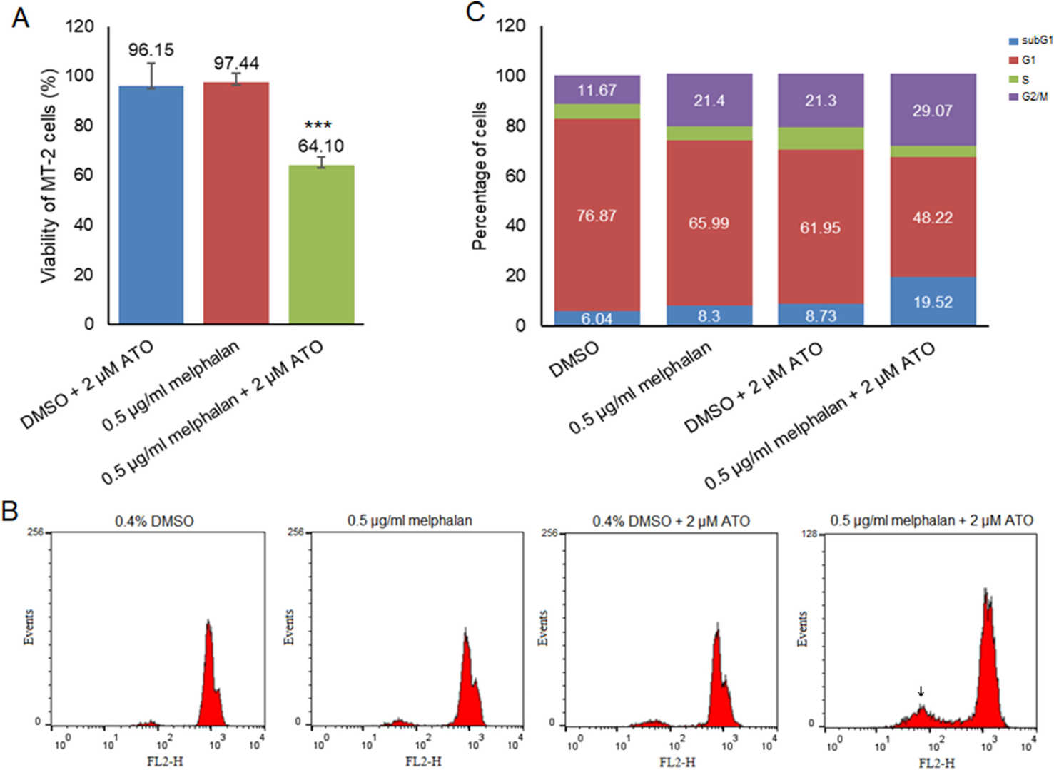 Fig. 1. Viability of MT-2 cells after 72 h treatment with melphalan and ATO, alone and in combination (A). Flow cytometry histograms (B) and quantitative analysis (C) of the cell cycle upon single and combinatorial use of melphalan and ATO (Khodadadi, Faeze, et al. 2025).
RELA/C Knockout Reduced FOXP3 Expression and Immune-suppressive Function of Tregs.
Mutations in NF-κB-related molecules result in combined immunodeficiency characterized by recurrent infection. This study aimed to investigate the association between mutations in NF-κB family members (RELA, RELB, and RELC) and regulatory T cell (Treg) function in humans. Similar to that observed in knockout mice, RELA or RELC knockout in MT-2 cells and freshly isolated Tregs reduced FOXP3 expression and the immune suppressive function of Tregs. Additionally, PD-L1 expression in effector T cells and Tregs decreased considerably following RELC knockdown. These findings demonstrated that the deletion of RELA or RELC resulted in the loss of Treg-like phenotype and function owing to the downregulation of FOXP3 expression.
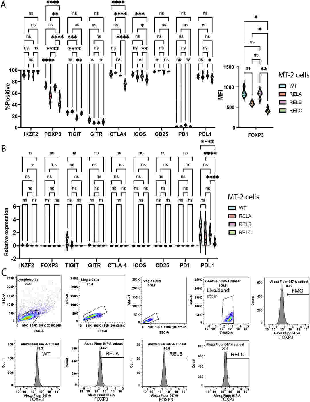 Fig. 2. FOXP3 expression was partially lost in MT-2 cells bearing RELA and RELC knockout (Sato, Yohei, Yamato Hanawa, and Akihito Tsubota. 2024).
Ask a Question
Write your own review
- You May Also Need

Description: Established in 2007 from the bone marrow mononuclear cells of an 82-year-old Japanese man with diffuse large B-cell lymphoma in the leukemic phase

Description: Established from the bone marrow of a 28-year-old man who developed the terminal leukemic phase of lymphosarcoma in 1976

Description: This cell line was derived from the bone marrow aspirate of a 59 year old male with erythroleukemia that became acute myelogenous leukaemia.The cells form colonies in soft-agar in the presence of ...

Description: Established from the pleural effusion of a 24-year-old woman with recurrent anaplastic large cell lymphoma (ALCL); cells were described to clonally derive from T-lineage lymphoid cells and to be ...

Description: Established from a 37-year-old man at second (refractory/terminal) relapse of Hodgkin lymphoma (nodular sclerosing -> lymphocyte depleted/stage IIISA -> stage IV) after both combined chemo- and ...

Description: Established from the peripheral blood of a 10-year-old Caucasian boy with acute lymphoblastic leukemia (pre B-ALL) at diagnosis in 1993
- Adipose Tissue-Derived Stem Cells
- Human Neurons
- Mouse Probe
- Whole Chromosome Painting Probes
- Hepatic Cells
- Renal Cells
- In Vitro ADME Kits
- Tissue Microarray
- Tissue Blocks
- Tissue Sections
- FFPE Cell Pellet
- Probe
- Centromere Probes
- Telomere Probes
- Satellite Enumeration Probes
- Subtelomere Specific Probes
- Bacterial Probes
- ISH/FISH Probes
- Exosome Isolation Kit
- Human Adult Stem Cells
- Mouse Stem Cells
- iPSCs
- Mouse Embryonic Stem Cells
- iPSC Differentiation Kits
- Mesenchymal Stem Cells
- Immortalized Human Cells
- Immortalized Murine Cells
- Cell Immortalization Kit
- Adipose Cells
- Cardiac Cells
- Dermal Cells
- Epidermal Cells
- Peripheral Blood Mononuclear Cells
- Umbilical Cord Cells
- Monkey Primary Cells
- Mouse Primary Cells
- Breast Tumor Cells
- Colorectal Tumor Cells
- Esophageal Tumor Cells
- Lung Tumor Cells
- Leukemia/Lymphoma/Myeloma Cells
- Ovarian Tumor Cells
- Pancreatic Tumor Cells
- Mouse Tumor Cells