
OVCAR-3
Cat.No.: CSC-C9120W
Species: Homo sapiens (Human)
Source: Ascites Metastasis
Morphology: epithelial
- Specification
- Background
- Scientific Data
- Q & A
- Customer Review
Established in 1982 from the malignant ascites of a patient with progressive ovarian adenocarcinoma following combination chemotherapy, OVCAR-3 is one of the most extensively characterized and widely utilized cell lines in ovarian cancer research. Bioinformatic and molecular analyses have firmly established OVCAR-3 as a faithful representative model of high-grade serous ovarian carcinoma (HGSOC), exhibiting the defining genomic hallmarks of the disease: functional loss of TP53, low somatic mutation burden, and high copy-number alteration frequency closely mirroring TCGA HGSOC profiles.
OVCAR-3 uniquely bridges chemoresistance and chemosensitivity phenotypes. While originally derived from a tumor clinically refractory to cisplatin, doxorubicin, and cyclophosphamide, the line demonstrates marked sensitivity to carboplatin and the PARP inhibitor rucaparib—comparable to BRCA-mutant models—attributed to functional homologous recombination repair deficiency. Concurrently, it harbors high non-homologous end-joining activity and amplifications in DNA damage response genes including ATR and TOPBP1, offering a sophisticated platform for dissecting DDR pathway dependencies and synthetic lethalities.
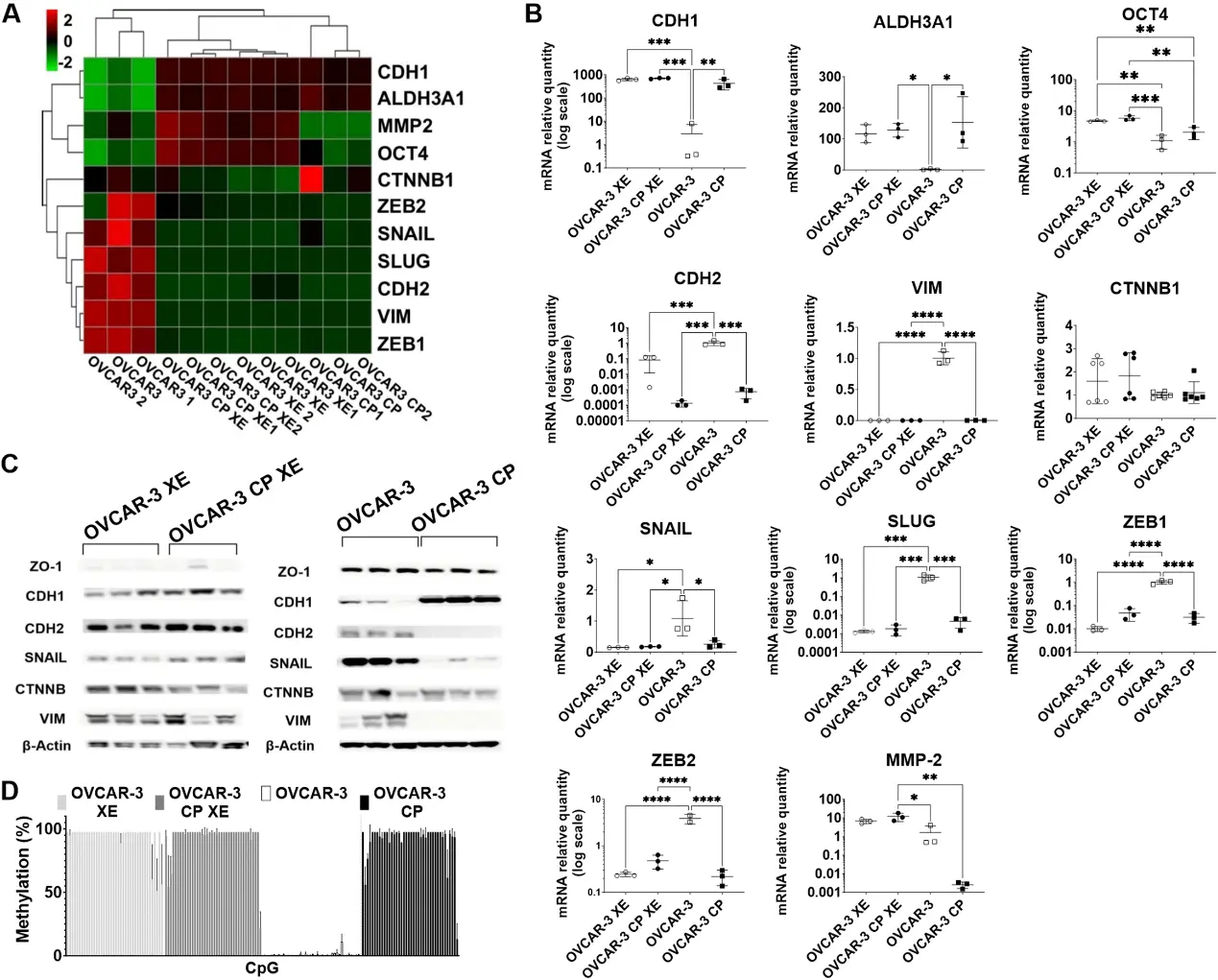
Stemness and EMT Profiles Shift in Xenografts Derived from Cisplatin-Sensitive and Cisplatin-Tolerant Ovarian Cancer Cells
The transcriptional alterations underlying epithelial-to-mesenchymal transition (EMT) in chemoresistant ovarian cancer remain a matter of debate, with emerging evidence pointing to tumor cell plasticity and subclone reprogramming.
In this study, Mrkvicova, Alena, et al. developed a cisplatin-tolerant ovarian cancer model by treating the cisplatin-sensitive OVCAR-3 cell line with a single dose of cisplatin, generating the OVCAR-3 CP variant. These cisplatin-tolerant cells exhibited distinct EMT-related changes at both transcriptomic and protein levels, potentially regulated by epigenetic mechanisms. EMT profiling revealed that OVCAR-3 CP cells did not display a pronounced mesenchymal phenotype but rather retained epithelial characteristics and showed elevated expression of ALDH3A1. In contrast, the parental chemosensitive OVCAR-3 cells expressed canonical mesenchymal markers (CDH2, VIM, ZEB1/2, SNAIL, SLUG) and lacked stemness marker expression. Upon xenografting, both OVCAR-3 and OVCAR-3 CP cells demonstrated phenotypic plasticity, with parental OVCAR-3 xenografts acquiring EMT-like features resembling to those observed in cisplatin-tolerant tumors. These findings suggest a decoupling of EMT from cisplatin-tolerance and instead underscore a stronger association between stemness traits and cisplatin tolerance.
Ask a Question
Write your own review
Description: Established from the malignant tumor of a 49-year-old woman with well-differentiated oxyphilic papillary thyroid carcinoma (PT2, G1, No, Mo); cells were described to be tumorigenic in nude mice; ...
Description: established from ovary of a 47-year-old female with ovarian endometrioid adenocarcinoma
Description: The A2780 human ovarian cancer cell line was established from tumour tissue from an untreated patient. Cells grow as a monolayer and in suspension in spinner cultures. A2780 is the parent line to the ...
Description: Japanese ovarian serous adenocarcinoma. Said CA125 producing. Cell growth is slow.
Description: Established post-hysterectomy from the tumor tissue of a 65-year-old Japanese woman with malignant ovarian carcinoma (stage: FIGO IIIc; histology: poorly differentiated serous papillary ...
- Adipose Tissue-Derived Stem Cells
- Human Neurons
- Mouse Probe
- Whole Chromosome Painting Probes
- Hepatic Cells
- Renal Cells
- In Vitro ADME Kits
- Tissue Microarray
- Tissue Blocks
- Tissue Sections
- FFPE Cell Pellet
- Probe
- Centromere Probes
- Telomere Probes
- Satellite Enumeration Probes
- Subtelomere Specific Probes
- Bacterial Probes
- ISH/FISH Probes
- Exosome Isolation Kit
- Human Adult Stem Cells
- Mouse Stem Cells
- iPSCs
- Mouse Embryonic Stem Cells
- iPSC Differentiation Kits
- Mesenchymal Stem Cells
- Immortalized Human Cells
- Immortalized Murine Cells
- Cell Immortalization Kit
- Adipose Cells
- Cardiac Cells
- Dermal Cells
- Epidermal Cells
- Peripheral Blood Mononuclear Cells
- Umbilical Cord Cells
- Monkey Primary Cells
- Mouse Primary Cells
- Breast Tumor Cells
- Colorectal Tumor Cells
- Esophageal Tumor Cells
- Lung Tumor Cells
- Leukemia/Lymphoma/Myeloma Cells
- Ovarian Tumor Cells
- Pancreatic Tumor Cells
- Mouse Tumor Cells