Glucocorticoid-Induced Osteoporosis (GIO) Model
If you are looking for a robust and reliable provider of disease models to support your osteoporosis research, Creative Bioarray is the perfect partner for you. Our glucocorticoid-induced osteoporosis (GIO) model is designed to meet the highest standards of scientific rigor and can provide you with the data you need to advance your research goals.
Glucocorticoids, commonly employed for their anti-inflammatory and immunosuppressive properties, are associated with a significant iatrogenic complication: GIO. Prolonged exposure to high doses of glucocorticoids leads to osteoporosis in a considerable number of patients. It is well-established that excess glucocorticoids directly impact bone cells, including osteoblasts, osteoclasts, and osteocytes, disrupting their proliferation, differentiation, apoptosis, and other vital functions. These cellular alterations are believed to play a crucial role in the pathogenesis of GIO.
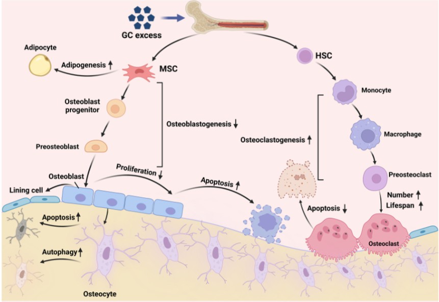 Fig. 1 Effects of GC excess on bone cell. GC, glucocorticoids
Our Glucocorticoid-Induced Osteoporosis (GIO) Model
- Available Animal
Rat - Modeling Method
Animals are treated with glucocorticoids (such as hydrocortisone or dexamethasone) to induce osteoporosis. - Endpoints
- Body weight
- Histology analysis: H&E staining
- Bone mineral density (BMD)
- Micro-CT
- Other customized endpoints
Example Data
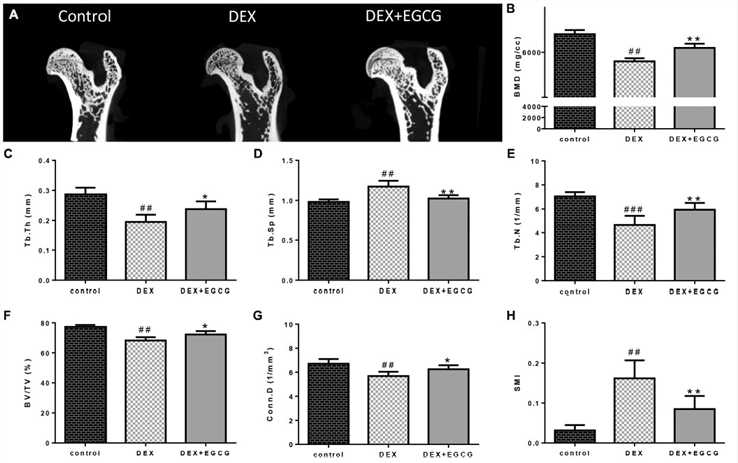 Fig. 2 Effects of EGCG on trabecular bone micro-architecture in GIO rats. (A) micro-CT of proximal femurs. (B) Data from BMD measurements of femurs by DXA. The following computed tomographic indices were analyzed in the defined region of interest (ROI): (C) Tb.Th, (D) Tb.Sp, (E) Tb.N, (F) BV/TV, (G) Conn.D, and (H) SMI.
Furthermore, we also provide other osteoporosis models that maybe you are interested in:
Quotation and Ordering
Creative Bioarray is dedicated to sharing our extensive expertise in drug efficacy studies with our clients to facilitate and accelerate their drug development process. We specialize in developing and validating disease models that mimic human pathologies, providing a rigorous testing environment for your drug candidates. If you are interested in our services, please feel free to contact us at any time or submit an inquiry to us directly. We look forward to cooperating with you.
References
- Chen, M., et al. Pathogenic mechanisms of glucocorticoid-induced osteoporosis. Cytokine & growth factor reviews, 2023, 70: 54-66.
- Liu, S., et al. Epigallocatechin-3-gallate ameliorates glucocorticoid-induced osteoporosis of rats in vivo and in vitro. Frontiers in pharmacology, 2018, 9: 447.
For research use only. Not for any other purpose.
Disease Models
- Oncology Models
-
Inflammation & Autoimmune Disease Models
- Rheumatoid Arthritis Models
- Glomerulonephritis Models
- Multiple Sclerosis (MS) Models
- Ocular Inflammation Models
- Sjögren's Syndrome Model
- LPS-induced Acute Lung Injury Model
- Peritonitis Models
- Passive Cutaneous Anaphylaxis Model
- Delayed-Type Hypersensitivity (DTH) Models
- Inflammatory Bowel Disease Models
- Systemic Lupus Erythematosus Animal Models
- Oral Mucositis Model
- Asthma Model
- Sepsis Model
- Psoriasis Model
- Atopic Dermatitis (AD) Model
- Scleroderma Model
- Gouty Arthritis Model
- Carrageenan-Induced Air Pouch Synovitis Model
- Carrageenan-Induced Paw Edema Model
- Experimental Autoimmune Myasthenia Gravis (EAMG) Model
- Graft-versus-host Disease (GvHD) Models
-
Cardiovascular Disease Models
- Surgical Models
- Animal Models of Hypertension
- Venous Thrombosis Model
- Atherosclerosis model
- Cardiac Arrhythmia Model
- Hyperlipoidemia Model
- Doxorubicin-induced Heart Failure Model
- Isoproterenol-induced Heart Failure Model
- Arterial Thrombosis Model
- Pulmonary Arterial Hypertension (PAH) Models
- Heart Failure with Preserved Ejection Fraction (HFpEF) Model
- Cardio-Renal-Metabolic (CKM) Syndrome Model
-
Neurological Disease Models
- Alzheimer's Disease Modeling and Assays
- Ischemic Stroke Models
- Acute Spinal Cord Injury (ASCI) Model
- Traumatic Brain Injury (TBI) Model
- Hypoxic-Ischemic Encephalopathy (HIE) Model
- Tourette Syndrome (TS) Model
- Amyotrophic Lateral Sclerosis (ALS) Model
- Huntington's Disease (HD) Model
- Intracerebral hemorrhage (ICH) Models
- Schizophrenia Model
- Depression Models
- Pain Models
-
Metabolic Disease Models
- Type 1 Diabetes Mellitus Model
- Type 2 Diabetes Mellitus Model
- Animal Model of Hyperuricemia
-
Nonalcoholic Fatty Liver Disease Model
- High-Fat Diet-Induced Nonalcoholic Fatty Liver Disease (NAFLD) Model
- Methionine and Choline Deficient (MCD) Diet-Induced Nonalcoholic Fatty Liver Disease (NAFLD) Model
- Gubra-Amylin NASH (GAN) Diet-Induced Nonalcoholic Fatty Liver Disease (NAFLD) Model
- Streptozotocin (STZ) Induced Nonalcoholic Fatty Liver Disease (NAFLD) Model
- High Fat Diet-Induced Obesity Model
- Diabetic Foot Ulcer (DFU) Model
- Liver Disease Models
- Rare Disease Models
- Respiratory Disease Models
- Digestive Disease Models
-
Urology Disease Models
- Cisplatin-induced Nephrotoxicity Model
- Unilateral Ureteral Obstruction Model
- 5/6 Nephrectomy Model
- Renal Ischemia-Reperfusion Injury (RIRI) Model
- Diabetic Nephropathy (DN) Models
- Passive Heymann Nephritis (PHN) Model
- Adenine-Induced Chronic Kidney Disease (CKD) Model
- Kidney Stone Model
- Doxorubicin-Induced Nephropathy Model
- Orthotopic Kidney Transplantation Model
- Benign Prostatic Hyperplasia (BPH) Model
- Peritoneal Fibrosis Model
- Orthopedic Disease Models
- Ocular Disease Models
- Infectious Disease Models
- Skin Disease Models
- Otology Disease Models