LPS-induced Acute Lung Injury Model
Acute lung injury (ALI) is a vital pulmonary inflammatory disorder with high mortality, and it can be caused by endotoxins, hyperoxia, etc. LPS (Lipopolysaccharide) -induced acute lung injury is a commonly utilized model that characterized by excessive inflammatory response within lung, hypoxemia and pulmonary oedema. LPS was shown to activate the expression of inflammatory mediators, such as tumor necrosis factor (TNF-α) and interleukin (IL)-6, by challenging mice/rats both intratracheally and intranasally. And subsequently, readouts are analyzed at specified time point.
Creative Bioarray focuses on drug research and development services and helps customers evaluate the drug efficacy and study the associated pathological mechanisms of ALI by LPS-induced acute lung injury model.
Our capabilities
- We screen novel test compounds targeting acute lung injury.
- We collect the BAL fluid of mice/rats from different groups and detect the expression of inflammatory mediators.
- We evaluate the MPO activity of lung tissues using MPO assay kit.
- We detect pathological changes of lung tissues between mice/rats from different groups.
Assays available
- BAL fluid collections
- Lung histology evaluation
- Cytokine analysis
- Lung oedema
- Total cell count
With extensive experience in the field of ALI, we are confident to help you to overcome any upcoming challenges. Our experts are fully capable of customizing our protocols and assays to meet your specific needs. With our help, we wish to facilitate your research with high efficiency.
Study examples
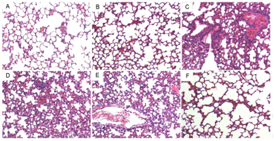 Figure. 1. Effects of alliin on histopathological changes in lung tissues in LPS-induced ALI mice. Representative histological changes of lung obtained from mice of different groups. A: Control group, B: alliin (100 mg/kg) alone group, C: LPS group, D: LPS+ alliin (25 mg/kg) group, E: LPS + alliin (50 mg/kg) group F: LPS + alliin (100 mg/kg) group (Hematoxylin and eosin staining, magnification 200×).
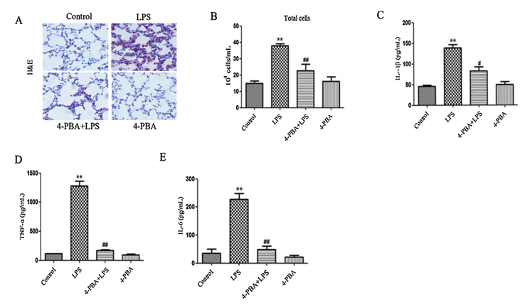 Figure. 2. Effect of 4-PBA on structural damage and production of inflammatory mediators induced by LPS in mice. (A) Lung sections of the mice were stained with representative H&E for histological examination (magnification × 200). (B) BAL fluid was performed to differentially count total cells. IL-1b (C), TNF-a (D) and IL-6 (E) in BAL fluid were analyzed by ELISA.
Quotation and ordering
If you have any special needs or questions regarding our services, please feel free to contact us. We look forward to cooperating with you in the future.
References
- Wang Y L, et al. Effects of alliin on LPS-induced acute lung injury by activating PPARγ[J]. Microbial Pathogenesis, 2017, 110:375-379.
- Zeng M, et al. 4-PBA inhibits LPS-induced inflammation through regulating ER stress and autophagy in acute lung injury models[J]. Toxicology Letters, 2017, 271(Complete):26-37.
For research use only. Not for any other purpose.
Disease Models
- Oncology Models
-
Inflammation & Autoimmune Disease Models
- Rheumatoid Arthritis Models
- Glomerulonephritis Models
- Multiple Sclerosis (MS) Models
- Ocular Inflammation Models
- Sjögren's Syndrome Model
- LPS-induced Acute Lung Injury Model
- Peritonitis Models
- Passive Cutaneous Anaphylaxis Model
- Delayed-Type Hypersensitivity (DTH) Models
- Inflammatory Bowel Disease Models
- Systemic Lupus Erythematosus Animal Models
- Oral Mucositis Model
- Asthma Model
- Sepsis Model
- Psoriasis Model
- Atopic Dermatitis (AD) Model
- Scleroderma Model
- Gouty Arthritis Model
- Carrageenan-Induced Air Pouch Synovitis Model
- Carrageenan-Induced Paw Edema Model
- Experimental Autoimmune Myasthenia Gravis (EAMG) Model
- Graft-versus-host Disease (GvHD) Models
-
Cardiovascular Disease Models
- Surgical Models
- Animal Models of Hypertension
- Venous Thrombosis Model
- Atherosclerosis model
- Cardiac Arrhythmia Model
- Hyperlipoidemia Model
- Doxorubicin-induced Heart Failure Model
- Isoproterenol-induced Heart Failure Model
- Arterial Thrombosis Model
- Pulmonary Arterial Hypertension (PAH) Models
- Heart Failure with Preserved Ejection Fraction (HFpEF) Model
- Cardio-Renal-Metabolic (CKM) Syndrome Model
-
Neurological Disease Models
- Alzheimer's Disease Modeling and Assays
- Seizure Models
- Parkinson's Disease Models
- Ischemic Stroke Models
- Acute Spinal Cord Injury (ASCI) Model
- Traumatic Brain Injury (TBI) Model
- Hypoxic-Ischemic Encephalopathy (HIE) Model
- Tourette Syndrome (TS) Model
- Amyotrophic Lateral Sclerosis (ALS) Model
- Huntington's Disease (HD) Model
- Intracerebral hemorrhage (ICH) Models
- Schizophrenia Model
- Depression Models
- Pain Models
-
Metabolic Disease Models
- Type 1 Diabetes Mellitus Model
- Type 2 Diabetes Mellitus Model
- Animal Model of Hyperuricemia
-
Nonalcoholic Fatty Liver Disease Model
- High-Fat Diet-Induced Nonalcoholic Fatty Liver Disease (NAFLD) Model
- Methionine and Choline Deficient (MCD) Diet-Induced Nonalcoholic Fatty Liver Disease (NAFLD) Model
- Gubra-Amylin NASH (GAN) Diet-Induced Nonalcoholic Fatty Liver Disease (NAFLD) Model
- Streptozotocin (STZ) Induced Nonalcoholic Fatty Liver Disease (NAFLD) Model
- High Fat Diet-Induced Obesity Model
- Diabetic Foot Ulcer (DFU) Model
- Cardio-Renal-Metabolic (CKM) Syndrome Model
- Liver Disease Models
- Rare Disease Models
- Respiratory Disease Models
- Digestive Disease Models
-
Urology Disease Models
- Cisplatin-induced Nephrotoxicity Model
- Unilateral Ureteral Obstruction Model
- 5/6 Nephrectomy Model
- Renal Ischemia-Reperfusion Injury (RIRI) Model
- Diabetic Nephropathy (DN) Models
- Passive Heymann Nephritis (PHN) Model
- Adenine-Induced Chronic Kidney Disease (CKD) Model
- Kidney Stone Model
- Doxorubicin-Induced Nephropathy Model
- Orthotopic Kidney Transplantation Model
- Benign Prostatic Hyperplasia (BPH) Model
- Peritoneal Fibrosis Model
- Cardio-Renal-Metabolic (CKM) Syndrome Model
- Orthopedic Disease Models
- Ocular Disease Models
- Infectious Disease Models
- Skin Disease Models
- Otology Disease Models