
SCC-9
Cat.No.: CSC-C9248W
Species: Homo sapiens (Human)
Source: Oral Cavity; Tongue
- Specification
- Background
- Scientific Data
- Q & A
- Customer Review
The SCC-9 cell line is a human squamous cell carcinoma cell line originally isolated from tongue carcinoma. SCC-9 cells are commonly used as an in vitro model for oral squamous cell carcinoma (OSCC). These cells are derived from malignant epithelial cells of the oral cavity and have been utilized in numerous studies concerning tumor biology, tumor invasion, and response to treatment in head and neck cancers.
Cultured SCC-9 cells adhere to the growth surface, showing a polygonal, epithelial-like appearance and forming compact colonies characterized by strong cell-cell junctions. Similar to other epithelial-derived and squamous cell carcinoma cells, SCC-9 cells express epithelial markers such as cytokeratins (CK13, CK14), E-cadherin, involucrin, and p63. Expression of epidermal growth factor receptor (EGFR) may also be present but is known to be variable. Dysregulation of EGFR has been shown to play a role in head and neck cancers.
Because of this, SCC-9 cells have been used to study many aspects of oral cancer including proliferation, differentiation, migration, invasion, and epithelial-mesenchymal transition (EMT). These cells are frequently used to study head and neck cancer related signaling pathways such as EGFR, PI3K/AKT, MAPK, and NF-κB and have been used to study mechanisms of tumor metastasis and chemoresistance/radiosensitivity. SCC-9 cells are also utilized for anticancer drug screening assays, radiosensitivity assays, and biomarker identification to aid in the development of targeted therapeutics for oral cancer and head and neck cancers.
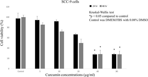
Effects of Curcumin on the Viability of SCC-9 Cells
In this study, Udompatankorn et al. investigated the proliferation and apoptosis of SCC-9 oral squamous cell carcinoma (OSCC) cells induced by curcumin and Lactobacillus rhamnosus GG cell-free supernatant (LGG CFS) both alone and in combination.
Curcumin decreased the viability of SCC-9 cells in a dose- and time-dependent manner. Curcumin concentrations ranging from 5 to 20 µg/ml did not markedly decrease the viability of SCC-9 cells after 24 and 48 h. However, treatment with 40 or 80 µg/ml curcumin decreased cell viability after 24 and 48 h (p < 0.05) (Fig. 1). Cell viabilities after treatment with 0, 5, 10, 20, 40, and 80 µg/ml curcumin for 24 h were 97.1%, 94.4%, 91.8%, 66.4%, 27.6%, and 28.2%, respectively, and similar results were obtained after 48 h. Therefore, we selected concentrations of 5 µg/ml (~5% inhibition, referred to as curcumin-L) and 40 µg/ml (>50% inhibition, referred to as curcumin-H) for additional studies with an incubation time of 24 h.
Ask a Question
Write your own review
Description: Human cell line derived from oral squamous cell carcinoma occurred in 69-yo, male patient. HLA-A 24/.

Description: Human cell line derived from metastasis of cancer occurred in oral cavity, the same patient as T3M-1 Clone2 and T3M-1 Cl-10.

Description: G-CSF and IL-1 producing oral squamous cell carcinoma, the same patient as T3M-1 Clone2 and CJM.

Description: The CAL-27 cells are established from the poorly differentiated squamous cell carcinoma of the tongue removed from a 56-year-old man before treatment in 1982. CAL 27 cells are epithelial, polygonal ...
Description: Established from the surgically removed fragment of a tongue lesion from a 69-year-old man with moderately differentiated squamous cell carcinoma of the tongue in 1983 (prior to therapy)

- Adipose Tissue-Derived Stem Cells
- Human Neurons
- Mouse Probe
- Whole Chromosome Painting Probes
- Hepatic Cells
- Renal Cells
- In Vitro ADME Kits
- Tissue Microarray
- Tissue Blocks
- Tissue Sections
- FFPE Cell Pellet
- Probe
- Centromere Probes
- Telomere Probes
- Satellite Enumeration Probes
- Subtelomere Specific Probes
- Bacterial Probes
- ISH/FISH Probes
- Exosome Isolation Kit
- Human Adult Stem Cells
- Mouse Stem Cells
- iPSCs
- Mouse Embryonic Stem Cells
- iPSC Differentiation Kits
- Mesenchymal Stem Cells
- Immortalized Human Cells
- Immortalized Murine Cells
- Cell Immortalization Kit
- Adipose Cells
- Cardiac Cells
- Dermal Cells
- Epidermal Cells
- Peripheral Blood Mononuclear Cells
- Umbilical Cord Cells
- Monkey Primary Cells
- Mouse Primary Cells
- Breast Tumor Cells
- Colorectal Tumor Cells
- Esophageal Tumor Cells
- Lung Tumor Cells
- Leukemia/Lymphoma/Myeloma Cells
- Ovarian Tumor Cells
- Pancreatic Tumor Cells
- Mouse Tumor Cells