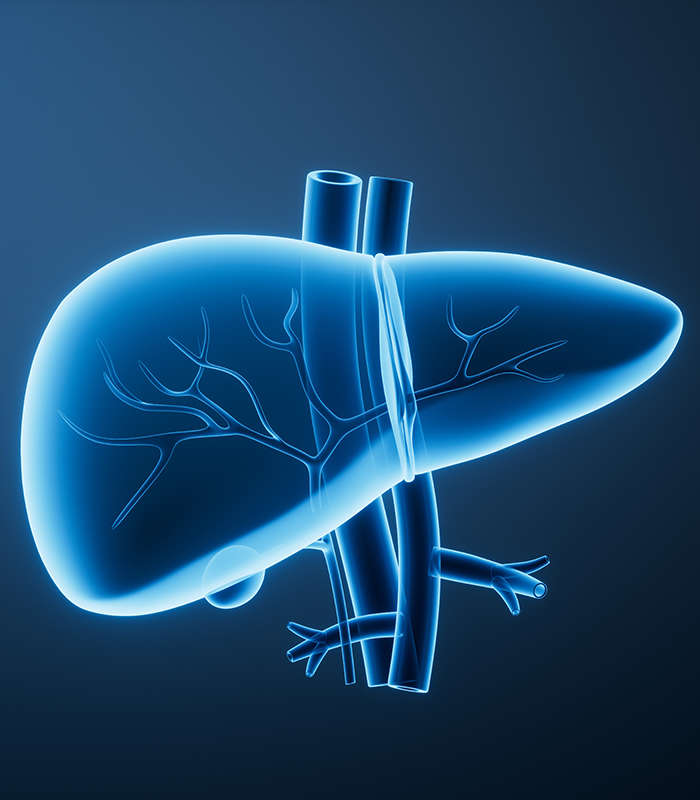
Gastric Tumor Cells
Gastric cancer is a highly heterogeneous malignancy with diverse histological and molecular profiles. As a leading cause of cancer-related mortality worldwide, understanding its complex biology—from H. pylori-induced transformation to advanced metastasis—requires physiologically relevant in vitro models.
Our Gastric Tumor Cell line collection encompasses a broad spectrum of models, including the classic Lauren classification (Intestinal and Diffuse types) and various molecular subtypes. These cell lines serve as essential tools for studying gastric mucosal barriers, epithelial-to-mesenchymal transition (EMT), and the development of targeted therapies.
High-Purity Phenotypically Diverse Well-Characterized Expert-Supported
Key Features & Expertise
Our gastric tumor cell lines are distinguished by their comprehensive biological relevance and rigorous validation:
Diverse Histological Representation
- Includes well, moderately, and poorly differentiated Gastric Adenocarcinomas.
- Specialized models for Signet Ring Cell Carcinoma and Mucinous Adenocarcinoma.
- Derived from various anatomical sites, including the antrum, body, and cardia, as well as metastatic sites like the liver and lymph nodes.
Advanced Molecular Profiling
- Defined HER2/neu expression levels for targeted drug screening and resistance studies.
- Models with documented EBV (Epstein-Barr Virus) and MSI (Microsatellite Instability) status.
- Genomic data available for key mutations, including TP53, CDH1 (E-cadherin), and PIK3CA.
Uncompromising Quality Control
- Authenticated via STR profiling to ensure genetic consistency and eliminate cross-contamination.
- Guaranteed Mycoplasma-free and tested for bacterial/fungal sterility.
- Optimized protocols provided for growth kinetics, media requirements, and cryopreservation.
FAQ
How do I choose the best gastric cancer cell line for HER2 research?
For studies involving HER2-targeted therapies (like Trastuzumab), NCI-N87 is a widely recognized model due to its high HER2 amplification. We provide specific expression data to help you select the most relevant model for your assay.
Do you provide normal gastric epithelial cells as experimental controls?
Yes. To ensure robust comparative data, we offer immortalized human gastric mucosal epithelial cell lines, which are essential for identifying tumor-specific changes in gene expression and drug response.
Are these cell lines suitable for studying drug resistance?
Absolutely. Our collection includes lines with varying sensitivities to standard chemotherapeutics like 5-Fluorouracil (5-FU), Cisplatin, and Taxanes. We also offer specific multidrug-resistant (MDR) variants for specialized research.
Can these cells be used for in vivo xenograft studies?
Many of our gastric tumor lines are highly tumorigenic and well-suited for subcutaneous or orthotopic xenograft models in nude mice, allowing for the translation of in vitro findings into complex biological systems.
What information is provided regarding the EBV status of the cell lines?
Since EBV-associated gastric cancer represents a distinct molecular subtype, we provide data on the EBV infection status for relevant lines, enabling researchers to study virus-host interactions in gastric carcinogenesis.
Filters Clear all filters
Species
- Cat (1)
- Human (989)
- Mouse (5)
- Rat (1)
Source
- Abdomen Metastasis (2)
- Adrenal Gland (7)
- Adrenal Gland Metastasis (2)
- Ascites (23)
- Ascites Metastasis (32)
- Bile Duct (3)
- Bladder (12)
- Blood (120)
- Bone (21)
- Bone Marrow (43)
- Bone Marrow Metastasis (18)
- Bone Metastasis (6)
- Brain (31)
- Brain Metastasis (6)
- Breast (8)
- Bronchus (1)
- Cecum (3)
- Cerebrospinal Fluid (1)
- Cerebrospinal Fluid Metastasis (1)
- Cervix (32)
- Colon (83)
- Cornea (3)
- Cutaneous Metastasis (1)
- Dermis (1)
- Duodenum (1)
- Endometrium (17)
- Esophagus (44)
- Eye (12)
- Eye Socket (5)
- Fetus (1)
- Foreskin (4)
- Gallbladder (1)
- Gingiva (2)
- Globe (2)
- Groin (1)
- Hypodermis Metastasis (5)
- Intestine (84)
- kidney (1)
- Kidney (9)
- Liver (13)
- Liver Metastasis (17)
- Lung (42)
- Lung Metastasis (8)
- Lymph Node (5)
- Lymph Node Metastasis (56)
- Muscle (4)
- Muscle Metastasis (2)
- Nose (2)
- Omentum Metastasis (2)
- Oral Cavity (10)
- Ovary (13)
- Ovary Metastasis (2)
- Pancreas (10)
- Pelvic Wall Metastasis (1)
- Pelvis (1)
- Perianal Space Metastasis (1)
- Pericardial Effusion (1)
- Pericardial Effusion Metastasis (1)
- Perineus (1)
- Peripheral Blood (119)
- Peritoneal Effusion (2)
- Peritoneum (1)
- Peritoneum Metastasis (1)
- Pharynx (3)
- Pleural Effusion (54)
- Pleural Effusion Metastasis (44)
- Prostate (4)
- Rectum (13)
- Renal Pelvis (1)
- Retroperitoneal Space (2)
- Salivary Gland (2)
- Skeletal Muscle (1)
- Skin (22)
- Skin Metastasis (3)
- Small Intestine (1)
- Small Intestine Metastasis (1)
- Soft Tissue Metastasis (1)
- Stomach (4)
- Testis (9)
- Thoracic Cavity Metastasis (6)
- Thyroid Gland (15)
- Thyroid Gland Metastasis (1)
- Tongue (5)
- Umbilical Cord (1)
- Umbilical Cord Blood (1)
- Urachus (1)
- Ureter (1)
- Uterus (53)
- Uvea (2)
- Vagina (2)
- Vulva (1)
Disease
- Acute Biphenotypic Leukemia (1)
- Acute Erythroid Leukemia (4)
- Acute Megakaryoblastic Leukemia (4)
- Acute Monocytic Leukemia (9)
- Acute Myeloid Leukemia (25)
- Acute Promyelocytic Leukemia (2)
- Adrenal Gland Neuroblastoma (11)
- Adult B Acute Lymphoblastic leukemia (1)
- Adult B Acute Lymphoblastic Leukemia (6)
- Adult T Acute Lymphoblastic Leukemia (6)
- Adult T Lymphoblastic Lymphoma (2)
- Adult T-Cell Leukemia/Lymphoma (1)
- Alveolar Rhabdomyosarcoma (4)
- Alveolar Ridge Squamous Cell Carcinoma (1)
- Amelanotic Melanoma (3)
- Ampulla of Vater Adenocarcinoma (1)
- Ampulla of Vater Adenosquamous Carcinoma (3)
- Anaplastic Astrocytoma (3)
- Anaplastic Large Cell Lymphoma (7)
- Askin Tumor (1)
- Astrocytoma (5)
- B Acute Lymphoblastic Leukemia (2)
- B-Cell Non-Hodgkin Lymphoma (5)
- Bare Lymphocyte Syndrome Type 2 (1)
- Barrett Adenocarcinoma (2)
- Benign Prostatic Hyperplasia (1)
- Bladder Carcinoma (12)
- Bladder Squamous Cell Carcinoma (1)
- Breast Adenocarcinoma (1)
- Breast Carcinoma (9)
- Breast Ductal Carcinoma (2)
- Burkitt Lymphoma (17)
- Canavan Disease (1)
- Cecum Adenocarcinoma (3)
- Central Nervous System Lymphoma (2)
- Cervical Adenocarcinoma (2)
- Cervical Adenosquamous Carcinoma (2)
- Cervical Small Cell Carcinoma (1)
- Cervical Squamous Cell Carcinoma (2)
- Childhood B Acute Lymphoblastic Leukemia (13)
- Childhood T Acute Lymphoblastic Leukemia (16)
- Childhood T Lymphoblastic Lymphoma (1)
- Cholangiocarcinoma (2)
- Chronic Eosinophilic Leukemia (1)
- Chronic Lymphocytic Leukemia (2)
- Chronic Myeloid Leukemia (23)
- Clear Cell Renal Cell Carcinoma (2)
- Colon Adenocarcinoma (53)
- Colon Carcinoma (33)
- Colorectal Adenocarcinoma (1)
- Colorectal Carcinoma (1)
- Congenital Pure Red Cell Aplasia (1)
- Cutaneous Melanoma (10)
- Dedifferentiated Chondrosarcoma (1)
- Desmoplastic Melanoma (1)
- Diffuse Large B-Cell Lymphoma (28)
- Down Syndrome (2)
- EBV-Related Burkitt Lymphoma (12)
- Embryonal Carcinoma (3)
- Embryonal Rhabdomyosarcoma (3)
- Endometrial Adenocarcinoma (13)
- Endometrial Adenosquamous Carcinoma (2)
- Endometrial Carcinoma (2)
- Endometrioid Stromal Sarcoma (1)
- Epithelioid Hemangioendothelioma (1)
- Epithelioid Sarcoma (3)
- Esophageal Adenocarcinoma (6)
- Esophageal Squamous Cell Carcinoma (41)
- Essential Thrombocythemia (1)
- Ewing Sarcoma (2)
- Extraskeletal Myxoid Chondrosarcoma (1)
- Fanconi Anemia (1)
- Fibrosarcoma (1)
- Follicular Lymphoma (2)
- Gallbladder Carcinoma (2)
- Gallbladder Undifferentiated Carcinoma (2)
- Gastric Adenocarcinoma (6)
- Gastric Adenosquamous Carcinoma (1)
- Gastric Carcinoma (5)
- Gastric Choriocarcinoma (1)
- Gastric Fundus Carcinoma (1)
- Gastric Signet Ring Cell Adenocarcinoma (1)
- Gastric Small Cell Carcinoma (2)
- Gastric Tubular Adenocarcinoma (5)
- Gastroesophageal Junction Adenocarcinoma (1)
- Gestational Choriocarcinoma (1)
- Gingival Squamous Cell Carcinoma (2)
- Glioblastoma (18)
- Gliosarcoma (1)
- Hairy Cell Leukemia (1)
- Hepatoblastoma (2)
- Hepatocellular Carcinoma (6)
- Hepatosplenic T-Cell Lymphoma (2)
- Hereditary Thyroid Gland Medullary Carcinoma (1)
- High Grade B-Cell Lymphoma (1)
- High Grade Ovarian Serous Adenocarcinoma (8)
- Hodgkin Lymphoma (9)
- Hypopharyngeal Squamous Cell Carcinoma (2)
- Infectious Mononucleosis (1)
- Intrahepatic Cholangiocarcinoma (6)
- Invasive Breast Carcinoma of No Special Type (12)
- Kidney Neoplasm (1)
- Kidney Rhabdoid Tumor (1)
- Krukenberg Tumor (1)
- Liposarcoma (1)
- Lung Adenocarcinoma (17)
- Lung Giant Cell Carcinoma (8)
- Lung Large Cell Carcinoma (9)
- Lung Mucoepidermoid Carcinoma (1)
- Lung Non-Small Cell Carcinoma (2)
- Lung Small Cell Carcinoma (25)
- Lung Squamous Cell Carcinoma (9)
- Lymphoblastic Lymphoma (1)
- Malignant Peripheral Nerve Sheath Tumor (1)
- Mantle Cell Lymphoma (5)
- Mature Gastric Teratoma (1)
- Maxillary Sinus Squamous Cell Carcinoma (1)
- Medulloblastoma (3)
- Melanoma (24)
- Meningioma (2)
- Minimally Invasive Lung Adenocarcinoma (1)
- Monophasic Synovial Sarcoma (1)
- Mouse Intestinal Tract Neuroendocrine Adenoma (1)
- Mouse Mammary Gland Malignant Neoplasm (2)
- Mouse Plasmacytoma (1)
- Mycosis Fungoides (1)
- Myelodysplastic Syndrome (1)
- Myxofibrosarcoma (1)
- Natural Killer Cell Lymphoblastic Leukemia/Lymphoma (2)
- Neuroblastoma (26)
- Oral Cavity Squamous Cell Carcinoma (15)
- Osteoid Osteoma (1)
- Osteosarcoma (15)
- Ovarian Carcinoma (1)
- Ovarian Clear Cell Adenocarcinoma (1)
- Ovarian Endometrioid Adenocarcinoma (4)
- Ovarian Granulosa Cell Tumor (1)
- Ovarian Mucinous Adenocarcinoma (2)
- Ovarian Serous Adenocarcinoma (2)
- Ovarian Serous Cystadenocarcinoma (2)
- Ovarian Small Cell Carcinoma (1)
- Pancreatic Adenocarcinoma (13)
- Pancreatic Carcinoma (5)
- Pancreatic Ductal Adenocarcinoma (12)
- Papillomavirus-Independent Cervical Squamous Cell Carcinoma (1)
- Papillomavirus-Related Cervical Adenocarcinoma (7)
- Papillomavirus-Related Cervical Squamous Cell Carcinoma (4)
- Papillomavirus-Related Endocervical Adenocarcinoma (16)
- Paroxysmal Nocturnal Hemoglobinuria (3)
- Pharyngeal Squamous Cell Carcinoma (1)
- Plasma Cell Myeloma (15)
- Pleural Epithelioid Mesothelioma (5)
- Pleural Sarcomatoid Mesothelioma (2)
- Poorly Differentiated Thyroid Gland Carcinoma (1)
- Primary Cutaneous T-Cell Non-Hodgkin Lymphoma (1)
- Primary Effusion Lymphoma (7)
- Primitive Neuroectodermal Tumor (1)
- Prostate carcinoma (1)
- Prostate Carcinoma (9)
- Rectal Adenocarcinoma (13)
- Rectosigmoid Adenocarcinoma (1)
- Recurrent Bladder Carcinoma (1)
- Renal Cell Carcinoma (7)
- Renal Pelvis Urothelial Carcinoma (1)
- Retinoblastoma (11)
- Sacral Chordoma (1)
- Sacrococcygeal Teratoma (1)
- Salivary Gland Squamous Cell Carcinoma (1)
- Sezary Syndrome (1)
- Shwachman-Diamond Syndrome (1)
- Skin Squamous Cell Carcinoma (2)
- Splenic Marginal Zone Lymphoma (1)
- Testicular Embryonal Carcinoma (8)
- Testicular Teratoma (2)
- Testicular Yolk Sac Tumor (1)
- Thyroid Gland Anaplastic Carcinoma (10)
- Thyroid Gland Follicular Carcinoma (4)
- Thyroid Gland Papillary Carcinoma (3)
- Thyroid Gland Sarcoma (1)
- Thyroid Gland Squamous Cell Carcinoma (2)
- Tongue Adenosquamous Carcinoma (1)
- Tongue Squamous Cell Carcinoma (6)
- Type I Endometrial Adenocarcinoma (1)
- Ureter Urothelial Carcinoma (1)
- Uterine Carcinosarcoma (2)
- Uterine Corpus Leiomyosarcoma (1)
- Uterine Corpus Sarcoma (2)
- Uveal Melanoma (2)
- Vaginal Melanoma (2)
- Vulvar Melanoma (1)
- Vulvar Squamous Cell Carcinoma (1)

Description: Species: human female 55 years old; Tissue: colon; Tumor: adenocarcinoma

Description: Species: human, Caucasian female 82 years old; Tissue: colon; Tumor: adenocarcinoma, grade IV

Description: Established from the poorly differentiated adenocarcinoma of the stomach (medullary type) of a ...

Description: The Cisplatin-resistant cell line MKN-45/DDP has been developed by repeatedly exposing the parent ...
Description: The Oxaliplatin-resistant cell line MKN-45/L-OHP has been developed by repeatedly exposing the ...

Description: Established in 1989 from the primary tumor of a 72-year-old Hispanic man with a poorly ...

Description: Species: human female 69 years; Tissue: colon; Tumor: carcinoma; Derived from: pelvic wall ...

Description: Human cell line derived from stomach cancer (gastric cancer; highly differentiated tubular ...

Description: Rectal carcinoma. Culture cell line of SCC.

Description: Human gastric cancer cell line derived from metastasis at liver.
Description: Human cell line derived from gastric cancer. Established from the cells in ascites.

Description: Human cell line derived from stomach cancer.

Description: Human cell line derived from signet ring cell carcinoma of stomach.
Description: Human cell line derived from intraperitoneal metastasis of gastric cancer

Description: Human cell line derived from gastric cancer

Description: Stomach cancer, but producing hybrid type of alkaline phosphatase.

Description: Stomach cancer, but producing Nagao-type alkaline phosphatase.

Description: Japanese gastric cancer, lymph node metastated.