Surgical Models
Cardiovascular disease is one of the risk factors affecting human health worldwide with increasing morbidity and mortality. The pathogenesis of heart disease is multifactorial and common causes of heart disease mainly consist of arterial hypertension, obesity, diabetes mellitus, renal dysfunction and inflammation. It is difficult to select a suitable model to investigate co-existing risk factors for certain cardiovascular disease.
Over the last few decades, surgical models, especially in rodents, have been widely used in the study of the pathomechanisms of cardiovascular disease due to their short breeding cycles and low housing costs. In addition, surgical techniques combined with genetic modifications and pharmacological approaches constitute a relatively comprehensive animal model.
Creative Bioarray specializes in providing customized pharmacodynamic research services to help customers assess the efficacy of drug candidates and study the associated pathological mechanisms through different surgical models.
Creative Bioarray provides you with surgical models including but not limitted to:
- Left coronary artery ligation model
- Transverse aortic constriction (TAC) model
- Ascending aortic constriction model
- Pulmonary artery constriction model
Species available
- Mouse
- Rat
- Guinea pig
Our Capabilities
- Left ventricular ejection fraction (LVEF), ventricular dimensions, and other indexes were detected by echocardiography.
- We detect changes in myocardial morphologyby H&E staining and so on.
- We evaluate myocardial ultrastructure by TEM.
Assays available
- Echocardiography
- Histopathological evaluation
- Biochemical analysis
- Transmission electron microscope (TEM)
With extensive experience in the field of cardiovascular disease, we are confident to help you overcome any upcoming challenges. Our experts are fully capable of customizing our protocols and assays to meet your specific needs. With our help, we wish to facilitate your research with high efficiency.
Study examples
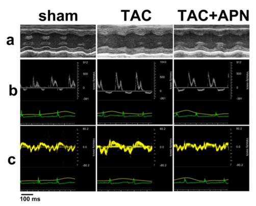 Figure. 1. Evaluation of cardiac function in three groups of mice. a M-mode echocardiography showed increased left ventricular posterior wall thickness (LVPW) in TAC mice while APNtreated mice demonstrated reduced LVPW. b Mitral inflow pattern and c mitral annular velocity of TAC group revealed progressive diastolic dysfunction while APN-treated mice showed recovery of diastolic dysfunction.
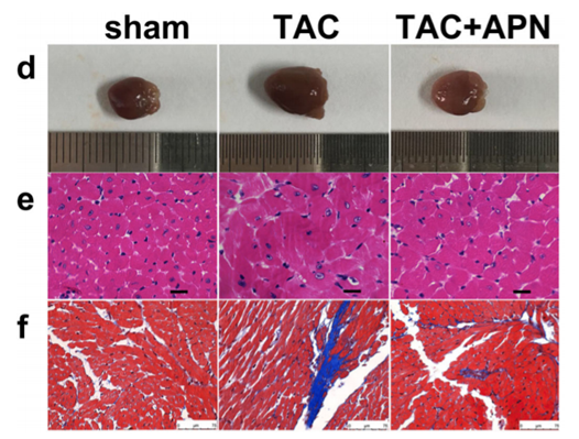 Figure. 2. Evaluation of cardiac structure, and morphology in three groups of mice. d Representative global heart photographs. e Hematoxylin and eosin-stained LV transverse sections (original magnification × 400; sale bar, 20 μm). f Representative microscopic images of Masson's trichrome staining (original magnification × 400; sale bar, 75 μm).
Quotation and ordering
If you have any special needs or questions regarding our services, please feel free to contact us. We look forward to cooperating with you in the future.
Reference
Han X, et al. Effects of Adiponectin on Diastolic Function in Mice Underwent Transverse Aorta Constriction[J]. Journal of Cardiovascular Translational Research, 2019:1-13.
For research use only. Not for any other purpose.
Disease Models
- Oncology Models
-
Inflammation & Autoimmune Disease Models
- Rheumatoid Arthritis Models
- Glomerulonephritis Models
- Multiple Sclerosis (MS) Models
- Ocular Inflammation Models
- Sjögren's Syndrome Model
- LPS-induced Acute Lung Injury Model
- Peritonitis Models
- Passive Cutaneous Anaphylaxis Model
- Delayed-Type Hypersensitivity (DTH) Models
- Inflammatory Bowel Disease Models
- Systemic Lupus Erythematosus Animal Models
- Oral Mucositis Model
- Asthma Model
- Sepsis Model
- Psoriasis Model
- Atopic Dermatitis (AD) Model
- Scleroderma Model
- Gouty Arthritis Model
- Carrageenan-Induced Air Pouch Synovitis Model
- Carrageenan-Induced Paw Edema Model
- Experimental Autoimmune Myasthenia Gravis (EAMG) Model
- Graft-versus-host Disease (GvHD) Models
-
Cardiovascular Disease Models
- Surgical Models
- Animal Models of Hypertension
- Venous Thrombosis Model
- Atherosclerosis model
- Cardiac Arrhythmia Model
- Hyperlipoidemia Model
- Doxorubicin-induced Heart Failure Model
- Isoproterenol-induced Heart Failure Model
- Arterial Thrombosis Model
- Pulmonary Arterial Hypertension (PAH) Models
- Heart Failure with Preserved Ejection Fraction (HFpEF) Model
- Cardio-Renal-Metabolic (CKM) Syndrome Model
-
Neurological Disease Models
- Alzheimer's Disease Modeling and Assays
- Seizure Models
- Parkinson's Disease Models
- Ischemic Stroke Models
- Acute Spinal Cord Injury (ASCI) Model
- Traumatic Brain Injury (TBI) Model
- Hypoxic-Ischemic Encephalopathy (HIE) Model
- Tourette Syndrome (TS) Model
- Amyotrophic Lateral Sclerosis (ALS) Model
- Huntington's Disease (HD) Model
- Intracerebral hemorrhage (ICH) Models
- Schizophrenia Model
- Depression Models
- Pain Models
-
Metabolic Disease Models
- Type 1 Diabetes Mellitus Model
- Type 2 Diabetes Mellitus Model
- Animal Model of Hyperuricemia
-
Nonalcoholic Fatty Liver Disease Model
- High-Fat Diet-Induced Nonalcoholic Fatty Liver Disease (NAFLD) Model
- Methionine and Choline Deficient (MCD) Diet-Induced Nonalcoholic Fatty Liver Disease (NAFLD) Model
- Gubra-Amylin NASH (GAN) Diet-Induced Nonalcoholic Fatty Liver Disease (NAFLD) Model
- Streptozotocin (STZ) Induced Nonalcoholic Fatty Liver Disease (NAFLD) Model
- High Fat Diet-Induced Obesity Model
- Diabetic Foot Ulcer (DFU) Model
- Cardio-Renal-Metabolic (CKM) Syndrome Model
- Liver Disease Models
- Rare Disease Models
- Respiratory Disease Models
- Digestive Disease Models
-
Urology Disease Models
- Cisplatin-induced Nephrotoxicity Model
- Unilateral Ureteral Obstruction Model
- 5/6 Nephrectomy Model
- Renal Ischemia-Reperfusion Injury (RIRI) Model
- Diabetic Nephropathy (DN) Models
- Passive Heymann Nephritis (PHN) Model
- Adenine-Induced Chronic Kidney Disease (CKD) Model
- Kidney Stone Model
- Doxorubicin-Induced Nephropathy Model
- Orthotopic Kidney Transplantation Model
- Benign Prostatic Hyperplasia (BPH) Model
- Peritoneal Fibrosis Model
- Cardio-Renal-Metabolic (CKM) Syndrome Model
- Orthopedic Disease Models
- Ocular Disease Models
- Infectious Disease Models
- Skin Disease Models
- Otology Disease Models