MRL/lpr Mouse Model
- Background
- Models
- Study Examples
- Features
- FAQ
To support in mechanistic studies and in vivo efficacy assessment, Creative Bioarray provides preclinical services for systemic lupus erythematosus (SLE) with the MRL/lpr mouse model. MRL/lpr mice, a popular model of lupus, spontaneously acquire autoimmune characteristics that closely mimic human illness, making them ideal for preclinical development and drug discovery.
With integrated readouts across functional, pathological, and biochemical endpoints, we enable robust assessment of therapeutic candidates in autoimmune disease models.
Background: Bridging the Gap in Lupus Research
Systemic lupus erythematosus is a complex autoimmune disease characterized by loss of immune tolerance and chronic systemic inflammation. It predominantly affects women and presents with multi-organ involvement, including kidney, skin, and central nervous system.
The pathogenesis of SLE involves a complex failure of immune tolerance. In the MRL/lpr model, a mutation in the Fas (CD95) gene that causes it to stop working makes lymphocyte apoptosis less effective. This causes an accumulation of self-reactive T and B cells, resulting in a huge generation of anti-dsDNA antibodies and antinuclear antibodies (ANA). These immune complexes accumulate in the kidneys and activate the inflammatory cascade, resulting in severe Lupus Nephritis (LN).
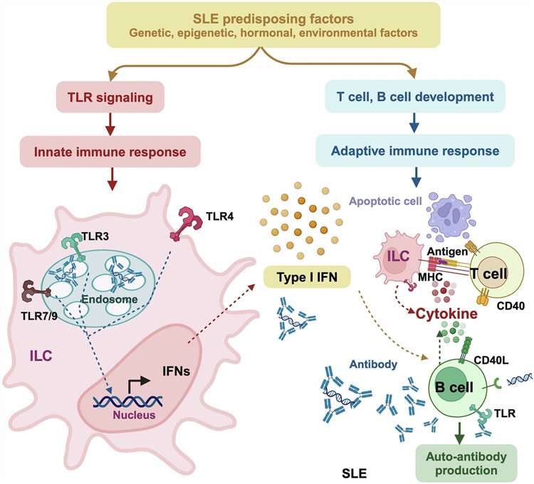 Fig. 1. Mechanisms-of-action driving SLE pathogenesis via over-activating immune response (Dai X, et al., 2025).
Corticosteroids and broad immunosuppressants are examples of current standard-of-care treatments that often result in serious side effects and poor long-term efficacy. As a result, before new precision therapies like BAFF inhibitors and IFN-pathway modulators are put into clinical trials, the MRL/lpr mouse model remains a widely used preclinical platform for evaluating the efficacy of novel therapeutic candidates prior to clinical translation.
Creative Bioarray's MRL/lpr Mouse Model
Strain: MRL/MpJ-Faslpr/J (MRL/lpr)
The MRL/lpr mouse carries a loss-of-function mutation in the Fas gene, leading to defective lymphocyte apoptosis. This results in:
- Accumulation of autoreactive lymphocytes
- Systemic autoimmunity
- Progressive lupus-like pathology
Key Endpoints:
- Serological & Autoimmune Markers: Anti-dsDNA, ANA, Total IgG, Cytokines (IL-6, TNF-α, CXCL13)
- Renal Function Biomarkers: Proteinuria (24 h urine), Blood Urea Nitrogen (BUN), Serum creatinine
- Renal Histopathology: H&E staining, PAS staining, etc
- Immunophenotyping: Flow cytometry for T/B cell activation (CD4+, CD8+, CD19+)
- Systemic Features: Kidney weight and kidney index, Spleen weight (splenomegaly), Lymph node weight
Study Examples
Lupus Mice Spontaneously Develop Splenomegaly
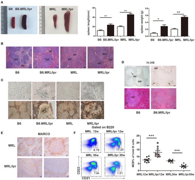 Fig. 2. MRL/lpr mice develop significant splenomegaly with enlarged spleen size and weight. Histology shows increased cellular accumulation and structural remodeling, along with impaired antigen capture function. Reduced MARCO⁺ macrophages and altered B cell populations further indicate disrupted splenic immune architecture (Zhang Q, et al. 2019).
Increase in pro-inflammatory immune cell subtypes
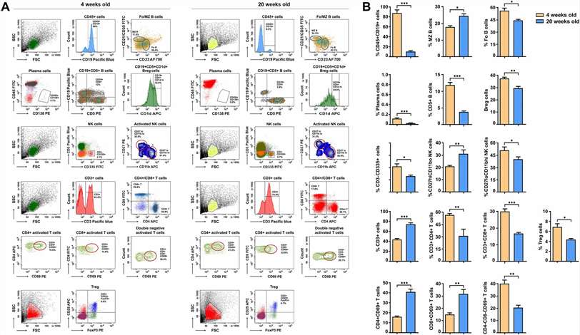 Fig. 3. Flow cytometry analysis shows a marked increase in pro-inflammatory immune cell subsets in aged MRL/lpr mice, including activated CD4⁺/CD8⁺ T cells and marginal zone B cells. In contrast, regulatory B and T cells are reduced, along with shifts in NK cell populations, indicating progressive immune dysregulation with disease progression (Delimitreva S, et al., 2024).
Elevation of IgG anti-dsDNA antibody levels and proteinuria
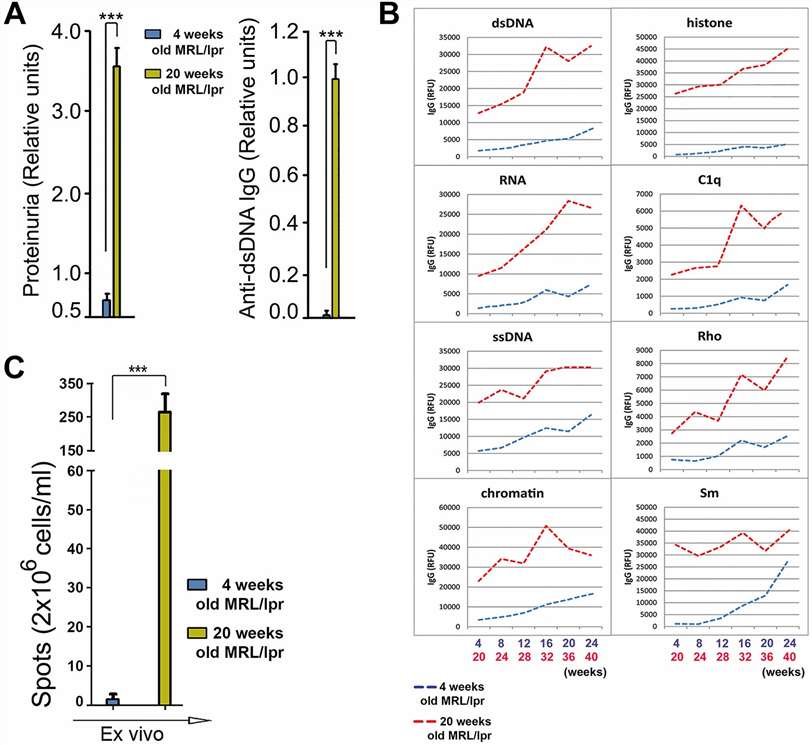 Fig. 4. Aged MRL/lpr mice show significantly elevated IgG anti-dsDNA antibody levels and progressive proteinuria, reflecting worsening lupus pathology (Delimitreva S, et al., 2024).
Why Choose Our MRL/lpr Model Services?

Integrated Readout Platforms
Combine immunology, pathology, and functional endpoints for comprehensive evaluation

Customizable Study Design
Flexible grouping, dosing regimens, and endpoint selection tailored to your drug

Translational Expertise
Strong alignment between preclinical endpoints and clinical biomarkers

Fast Turnaround
Large-scale colonies ready for immediate study enrollment

Data Quality and Reproducibility
Standardized protocols and rigorous QC ensure consistent results
FAQ
What is the MRL/lpr mouse model?
The MRL/lpr mouse model is a spontaneous autoimmune model carrying a Fas mutation, widely used to study systemic lupus erythematosus.
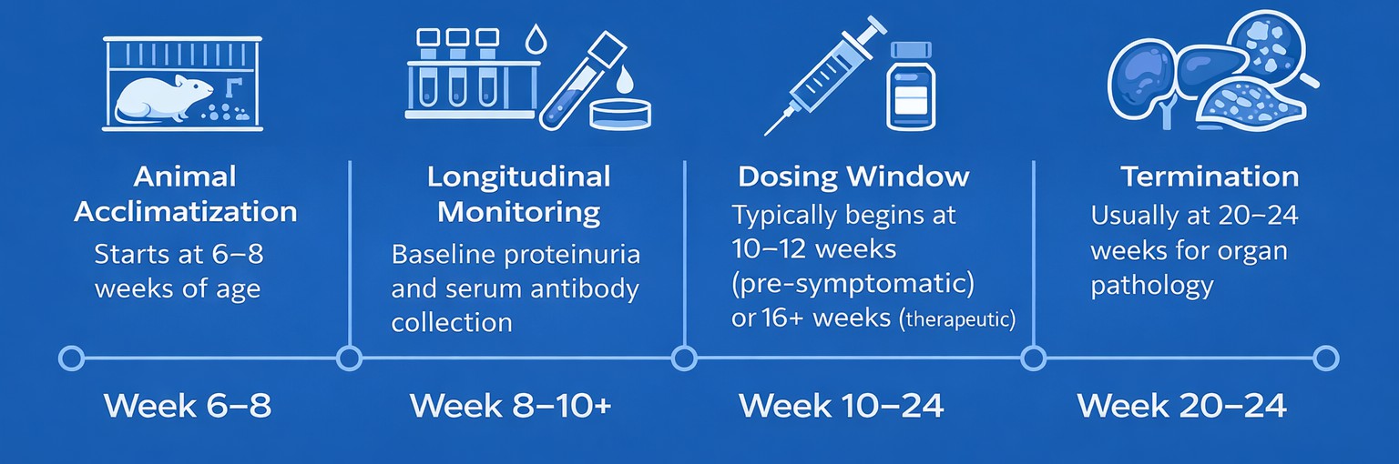
What is the typical onset of Lupus Nephritis in this model?
While antibodies appear at 12 weeks, significant proteinuria and histological kidney damage typically stabilize between 16 and 20 weeks.
Are there gender differences in disease severity?
Yes, female MRL/lpr mice generally exhibit earlier onset and more aggressive disease progression than males.
How does the MRL/lpr model compare to NZB/W mice?
MRL/lpr mice develop disease earlier and more aggressively, while NZB/W mice show slower disease progression, suitable for chronic studies.
What endpoints are commonly used in lupus drug testing mouse models?
Common endpoints include proteinuria, anti-dsDNA antibodies, cytokine levels, and kidney histopathology.
Start Your Lupus Preclinical Study
Accelerate your SLE drug development with a validated MRL/lpr mouse model.
Creative Bioarray offers end-to-end CRO services, from study design to data analysis, supporting your preclinical efficacy evaluation and translational research.
References
- Dai X, Fan Y, et al. Systemic lupus erythematosus: updated insights on the pathogenesis, diagnosis, prevention and therapeutics. Sig Transduct Target Ther. 2025. 10, 102.
- Zhang Q, Xiang L, et al. Predominant Role of Immunoglobulin G in the Pathogenesis of Splenomegaly in Murine Lupus. Front Immunol. 2020. 10:3020.
- Delimitreva S, Boneva G, et al. Lupus progression deteriorates oogenesis quality in MRL/lpr mice. Immunol Res. 2024. 72, 811–827