Spontaneous Arthritis Models
Rheumatoid arthritis (RA) is affected by the interaction of both genetic and environmental factors. Unfortunately, no model organisms could accurately simulate all pathological characteristics of RA at present. Creative Bioarray focuses on drug research and development services and helps customers assess the drug efficacy against RA and study the associated pathological mechanisms of RA.
Spontaneous Arthritis Models include but not limited to:
- K/BxN spontaneous mouse model
- TNF-α transgenic mouse model
- SKG model
- Human DR4-CD4 mice
- Human/SCID chimeric mice
Our capabilities
- We use Spontaneous Arthritis Models to test the efficacy of drug candidates targeting Rheumatoid arthritis.
- We assess the severity of rheumatoid arthritis in animals by Clinical scoring system.
- We evaluate various biomarkers and cytokines through WB, IHC, ELISA, etc.
Assays available
- Clinical score
- Histopathology
- Biomarker analysis
- Cytokine analysis
- PK/PD blood analysis
- Paw volume
- Ankle thickness
With extensive experience in the field of RA, we are confident to help you to overcome any upcoming challenges. Our experts are fully capable of customizing our protocols and assays to meet your specific needs. With our help, we wish to facilitate your research with high efficiency.
Study examples
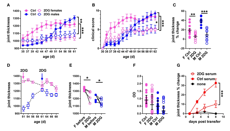 Figure. 1. Glycolysis inhibition reduced joint inflammation in KBN mice. Time course of joint thickness (A) and clinical scores (B) in mice treated or not with 2DG (female controls N = 11; female 2DG N = 12; male controls N = 7; male 2DG N = 9). (C). Percent change in joint thickness between d 35 and d 61 in these mice (t-tests). (D). Time course of joint thickness in mice in which treatment with 2DG started after severe joint inflammation was established, at 51 d old for females and 61 d old for males (as indicated by arrows). (E). Changes in joint thickness after 2DG treatment shown in (D) in individual mice (initial and terminal measurements, paired t-tests). (F). Serum anti-GPI IgG in mice treated preventively or not with 2DG as shown in (A,B) (geometric means ± 95% confidence intervals). (G). Joint thickness in KRN mice after transfer of serum from KBN mice treated with 2DG or controls (N = 6 each). An uninjected KRN mouse is included as control. Plots in (A,B,G) were compared by 2-way ANOVA. *p < 0.05; **p < 0.01; ***p < 0.001.
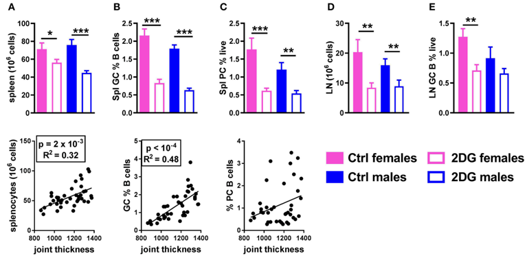 Figure. 2. Glycolysis inhibition reduced lymphoid expansion as well as GC B cells and plasma cell differentiation. Mice treated with 2DG showed a reduced number of splenocytes (A) and frequency of GL7+CD95+ GC B cells (B), both of which were correlated with disease severity, as well as a reduction of the frequency of CD138+B220lo/neg plasma cells, which was not correlated with disease activity (C). 2DG also reduced the number of cells (D) and the frequency of GC B cells (E) in the JDLN. Mean values between the treated and control mice were evaluated with t-tests. *p < 0.05; **p < 0.01; ***p < 0.001. Correlations between joint thickness and immune variables were computed by pooling the 4 groups of mice and evaluated with a Pearson test as indicated for each graph.
Quotation and ordering
If you have any special needs or questions regarding our services, please feel free to contact us. We look forward to cooperating with you in the future.
Reference
Auci D L et al. Oral treatment with HE3286 ameliorates disease in rodent models of rheumatoid arthritis[J]. International Journal of Molecular Medicine, 2010, 25(4):625-633.