Neurological Disease Models

It is well known that central nervous system (CNS) diseases are the leading cause of the global disease burden. However, although advances in fundamental neuroscience are rapidly accelerating, our understanding of brain function and dysfunction has limited impact on new therapies and treatments for central nervous system diseases. This may be partly because researchers still face a fundamental unanswered question: What constitutes a "good" animal or cell model of neurological disease?
It is indispensable and effective to use animal disease models in experimental medical research. Animal models of neurological disease provide a convenient and reliable platform for developing new therapies and drugs for such diseases. Creative Bioarray uses animal models of different neurological diseases to provide professional drug or therapy testing and research services.
The induction methods of rodent neurological disease models include chemical drugs, surgical induction, and genetic modification. Our pharmacology team has developed a variety of neurological disease models based on the needs of our customers for new drugs or therapy testing.
Our portfolio of neurological disease models cover the following diseases
- Alzheimer's disease
- Seizure
- Parkinson's disease
- Ischemic Stroke Model
- Acute Spinal Cord Injury (ASCI) Model
- Traumatic Brain Injury (TBI) Model
- Hypoxic-Ischemic Encephalopathy (HIE) Model
- Tourette Syndrome (TS) Model
- Amyotrophic Lateral Sclerosis (ALS) Model
- Huntington's Disease Model
- Intracerebral Hemorrhage (ICH) Models
- Schizophrenia Model
- Depression Models
We provide highly customizable services
- Customized animal feeding
- Customized samples collecting
- Biomarker analysis
- Detection of multiple proteins and cytokines
- RNA detection and analysis
- High-throughput sequencing analysis
- Pathology testing
- Immunohistochemistry
Study example
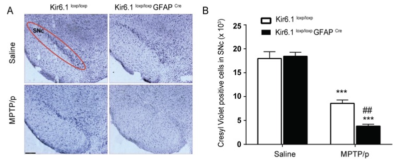
 Figure 1. Astrocytic Kir6.1 knockout exacerbates the loss of DA neurons and promotes astrocyte over-activation in SNc of MPTP/p PD model mice. A, Microphotographs of Cresyl violet-positive cells in the substantia nigra compacta (SNc). B, Stereological counts of Cresyl violet-positive cells in the SNc(Hu et al., 2019).
Additionally, our professionals have the expertise and research equipment to study the interaction between the microbiome and metabolic diseases, which attribute critical new aspects to customers' drug development research.
Quotation and ordering
We have extensive experience in developing disease models based on scientific publications. To discuss any of these models further or to discuss the possibility of developing alternative models, please do not hesitate to contact us.
Reference
- Hu, Z.-L.; et al. Kir6.1/K-ATP channel on astrocytes protects against dopaminergic neurodegeneration in the MPTP mouse model of Parkinson's disease via promoting mitophagy. Brain, Behavior, and Immunity, (2019), 81, 509–522.
For research use only. Not for any other purpose.
Disease Models
- Oncology Models
-
Inflammation & Autoimmune Disease Models
- Rheumatoid Arthritis Models
- Glomerulonephritis Models
- Multiple Sclerosis (MS) Models
- Ocular Inflammation Models
- Sjögren's Syndrome Model
- LPS-induced Acute Lung Injury Model
- Peritonitis Models
- Passive Cutaneous Anaphylaxis Model
- Delayed-Type Hypersensitivity (DTH) Models
- Inflammatory Bowel Disease Models
- Systemic Lupus Erythematosus Animal Models
- Oral Mucositis Model
- Asthma Model
- Sepsis Model
- Psoriasis Model
- Atopic Dermatitis (AD) Model
- Scleroderma Model
- Gouty Arthritis Model
- Carrageenan-Induced Air Pouch Synovitis Model
- Carrageenan-Induced Paw Edema Model
- Experimental Autoimmune Myasthenia Gravis (EAMG) Model
- Graft-versus-host Disease (GvHD) Models
-
Cardiovascular Disease Models
- Surgical Models
- Animal Models of Hypertension
- Venous Thrombosis Model
- Atherosclerosis model
- Cardiac Arrhythmia Model
- Hyperlipoidemia Model
- Doxorubicin-induced Heart Failure Model
- Isoproterenol-induced Heart Failure Model
- Arterial Thrombosis Model
- Pulmonary Arterial Hypertension (PAH) Models
- Heart Failure with Preserved Ejection Fraction (HFpEF) Model
- Cardio-Renal-Metabolic (CKM) Syndrome Model
-
Neurological Disease Models
- Alzheimer's Disease Modeling and Assays
- Seizure Models
- Parkinson's Disease Models
- Ischemic Stroke Models
- Acute Spinal Cord Injury (ASCI) Model
- Traumatic Brain Injury (TBI) Model
- Hypoxic-Ischemic Encephalopathy (HIE) Model
- Tourette Syndrome (TS) Model
- Amyotrophic Lateral Sclerosis (ALS) Model
- Huntington's Disease (HD) Model
- Intracerebral hemorrhage (ICH) Models
- Schizophrenia Model
- Depression Models
- Pain Models
-
Metabolic Disease Models
- Type 1 Diabetes Mellitus Model
- Type 2 Diabetes Mellitus Model
- Animal Model of Hyperuricemia
-
Nonalcoholic Fatty Liver Disease Model
- High-Fat Diet-Induced Nonalcoholic Fatty Liver Disease (NAFLD) Model
- Methionine and Choline Deficient (MCD) Diet-Induced Nonalcoholic Fatty Liver Disease (NAFLD) Model
- Gubra-Amylin NASH (GAN) Diet-Induced Nonalcoholic Fatty Liver Disease (NAFLD) Model
- Streptozotocin (STZ) Induced Nonalcoholic Fatty Liver Disease (NAFLD) Model
- High Fat Diet-Induced Obesity Model
- Diabetic Foot Ulcer (DFU) Model
- Cardio-Renal-Metabolic (CKM) Syndrome Model
- Liver Disease Models
- Rare Disease Models
- Respiratory Disease Models
- Digestive Disease Models
-
Urology Disease Models
- Cisplatin-induced Nephrotoxicity Model
- Unilateral Ureteral Obstruction Model
- 5/6 Nephrectomy Model
- Renal Ischemia-Reperfusion Injury (RIRI) Model
- Diabetic Nephropathy (DN) Models
- Passive Heymann Nephritis (PHN) Model
- Adenine-Induced Chronic Kidney Disease (CKD) Model
- Kidney Stone Model
- Doxorubicin-Induced Nephropathy Model
- Orthotopic Kidney Transplantation Model
- Benign Prostatic Hyperplasia (BPH) Model
- Peritoneal Fibrosis Model
- Cardio-Renal-Metabolic (CKM) Syndrome Model
- Orthopedic Disease Models
- Ocular Disease Models
- Skin Disease Models
- Infectious Disease Models
- Otology Disease Models