Orchiectomy (ORX)-Induced Osteoporosis Model
Creative Bioarray is proud to offer our clients an advanced and reliable orchiectomy (ORX)-induced osteoporosis model. This model is specifically designed to help evaluate the efficacy of drugs and study the underlying mechanism of osteoporosis. With our state-of-the-art technology and expertise, we are committed to providing our clients with the highest quality research solutions to meet their specific needs.
Osteoporosis has long been considered a disease that mainly affects post-menopausal women. However, many studies have shown that elderly men are also at risk of developing this condition. Male osteoporosis is associated with several risk factors, including androgen deficiency. In animal models, ORX has been shown to result in decreased bone mass, particularly in rodents, with a decrease of BMD and BV/TV and microarchitecture alterations due to an increased bone remodeling. Therefore, the ORX animal model is widely used in male osteoporosis research.
Our Orchiectomy (ORX)-Induced Osteoporosis Model
- Available Animal
Rat - Modeling Method
After anesthetization, animals are induced osteoporosis by orchiectomy. - Endpoints
- Body weight
- Histology analysis: H&E staining
- Bone mineral density (BMD)
- Micro-CT
- Serum analysis: osteocalcin, alkaline phosphatase (ALP), etc.
- Other customized endpoints
Example Data
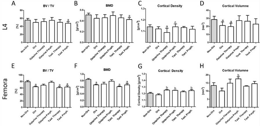 Fig. 1 3D micro-CT analysis. 3D analysis of L4 (A–D) and femur (E–H) performed after 18 weeks in Non-Orx rats and Orx rats either untreated or treated with ostarine or testosterone.
Moreover, we also provide other osteoporosis models that maybe you are interested in:
Quotation and Ordering
Creative Bioarray is a leading provider of disease models for drug pre-clinical research. With years of experience, we have developed a wide range of models that can help accelerate drug discovery and development. Our team of experts is always willing to share their knowledge and expertise in pharmacology with our clients, ensuring they have the support they need to succeed in their research endeavors. If you are interested in our services, please feel free to contact us at any time or submit an inquiry to us directly. We look forward to cooperating with you.
References
- Böker, K.O., et al. Treatment of osteoporosis using a selective androgen receptor modulator ostarine in an orchiectomized rat model. Endocrine, 2023, 81(3): 579-591.
- Blouin, S., et al. Orchidectomy models of osteoporosis. Osteoporosis: Methods and Protocols, 2008: 125-134.
For research use only. Not for any other purpose.
Disease Models
- Oncology Models
-
Inflammation & Autoimmune Disease Models
- Rheumatoid Arthritis Models
- Glomerulonephritis Models
- Multiple Sclerosis (MS) Models
- Ocular Inflammation Models
- Sjögren's Syndrome Model
- LPS-induced Acute Lung Injury Model
- Peritonitis Models
- Passive Cutaneous Anaphylaxis Model
- Delayed-Type Hypersensitivity (DTH) Models
- Inflammatory Bowel Disease Models
- Systemic Lupus Erythematosus Animal Models
- Oral Mucositis Model
- Asthma Model
- Sepsis Model
- Psoriasis Model
- Atopic Dermatitis (AD) Model
- Scleroderma Model
- Gouty Arthritis Model
- Carrageenan-Induced Air Pouch Synovitis Model
- Carrageenan-Induced Paw Edema Model
- Experimental Autoimmune Myasthenia Gravis (EAMG) Model
- Graft-versus-host Disease (GvHD) Models
-
Cardiovascular Disease Models
- Surgical Models
- Animal Models of Hypertension
- Venous Thrombosis Model
- Atherosclerosis model
- Cardiac Arrhythmia Model
- Hyperlipoidemia Model
- Doxorubicin-induced Heart Failure Model
- Isoproterenol-induced Heart Failure Model
- Arterial Thrombosis Model
- Pulmonary Arterial Hypertension (PAH) Models
- Heart Failure with Preserved Ejection Fraction (HFpEF) Model
- Cardio-Renal-Metabolic (CKM) Syndrome Model
-
Neurological Disease Models
- Alzheimer's Disease Modeling and Assays
- Ischemic Stroke Models
- Acute Spinal Cord Injury (ASCI) Model
- Traumatic Brain Injury (TBI) Model
- Hypoxic-Ischemic Encephalopathy (HIE) Model
- Tourette Syndrome (TS) Model
- Amyotrophic Lateral Sclerosis (ALS) Model
- Huntington's Disease (HD) Model
- Intracerebral hemorrhage (ICH) Models
- Schizophrenia Model
- Depression Models
- Pain Models
-
Metabolic Disease Models
- Type 1 Diabetes Mellitus Model
- Type 2 Diabetes Mellitus Model
- Animal Model of Hyperuricemia
-
Nonalcoholic Fatty Liver Disease Model
- High-Fat Diet-Induced Nonalcoholic Fatty Liver Disease (NAFLD) Model
- Methionine and Choline Deficient (MCD) Diet-Induced Nonalcoholic Fatty Liver Disease (NAFLD) Model
- Gubra-Amylin NASH (GAN) Diet-Induced Nonalcoholic Fatty Liver Disease (NAFLD) Model
- Streptozotocin (STZ) Induced Nonalcoholic Fatty Liver Disease (NAFLD) Model
- High Fat Diet-Induced Obesity Model
- Diabetic Foot Ulcer (DFU) Model
- Liver Disease Models
- Rare Disease Models
- Respiratory Disease Models
- Digestive Disease Models
-
Urology Disease Models
- Cisplatin-induced Nephrotoxicity Model
- Unilateral Ureteral Obstruction Model
- 5/6 Nephrectomy Model
- Renal Ischemia-Reperfusion Injury (RIRI) Model
- Diabetic Nephropathy (DN) Models
- Passive Heymann Nephritis (PHN) Model
- Adenine-Induced Chronic Kidney Disease (CKD) Model
- Kidney Stone Model
- Doxorubicin-Induced Nephropathy Model
- Orthotopic Kidney Transplantation Model
- Benign Prostatic Hyperplasia (BPH) Model
- Peritoneal Fibrosis Model
- Orthopedic Disease Models
- Ocular Disease Models
- Infectious Disease Models
- Skin Disease Models
- Otology Disease Models